X-ray Imaging
The articles presented in this chapter illustrate the broad range of scientific topics encountered at the X-ray imaging beamlines, from medicine and life science to palaeontology, nanowires, stardust or historical artefacts. All the examples share a common property: the heterogeneity (and often complexity) of the samples. However, they usually differ in terms of size, composition and structure. While ID17, the biomedical beamline, offers a beam large enough to image objects the size of organs (for example Figure 49, imaging the complete human knee), the NINA project (nano-imaging and nano-analysis, upgrade beamline project UPBL4) provides two new beamlines, ID16A and ID16B, designed to deliver the ultimate nanometric resolution. Accordingly, the five X-ray imaging beamlines offer our users a very wide range of accessible lateral resolution and corollary sample sizes, as well as an extended set of imaging and analytical tools.
 |
|
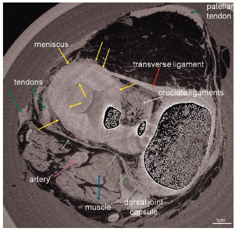
Fig. 49: Axial section of a full human knee in vitro imaged at ID17 using propagation-based phase contrast imaging. The image reveals an unprecedented visualisation of the various soft tissue structures (from [1]). |
This year, the NINA project has reached fruition. ID16B, the nano-analysis beamline, welcomed its first users in April. Two beam modes can be used: pink beam and monochromatic beam and various techniques are available using a nanobeam focused down to around 50 × 50 nm2: 2D and 3D fluorescence, 2D and 3D diffraction, 3D phase contrast imaging and 2D X-ray excited optical emission (XEOL). The very first papers based on these pioneering experiments have recently been accepted. As an example, Laforce et al. reports, the 3D nano fluorescence imaging of meteoric nanoparticles in the journal Analytical Chemistry [2]. Nano-X-ray absorption spectroscopy (XAS), one important deliverable of the beamline project, still requires additional development and commissioning, but should be available in 2015. At ID16A, the nano-imaging beamline, the first users arrived in October. They were very pleased to test a focused beam as small as 35 nm vertical by 20 nm horizontal at 17 keV with a flux as high as 1012 photons/s. The two main imaging configurations, X-ray fluorescence analysis and magnified phase tomography, are now regularly being put to good use, either on their own or in combination during a single experiment. The beamline will add three major capabilities in 2015: a high energy focusing mirror operating at 34 keV, ptychographic imaging for ultimate resolution and cryogenic sample preservation.
At an intermediate submicrometric scale, ID21, the spectromicroscopy beamline, offers complementary tools to the NINA beamlines with an X-ray microscope in the tender X-ray domain (2-9 keV) for 2D X-ray fluorescence (XRF) and XAS, a FTIR microscope for molecular mapping, including a side branch that should soon provide users with a stable and easy to use instrument for 2D µXRF and µXRD mapping. This is currently the main project at ID21, which is being implemented without any impact on the regular operation of the main branch. In 2014, major achievements included producing the first monochromatic beam in July 2014 (fixed energy at 8.5 keV) and the first focused beam in November 2014 (0.9 µm vertical × 2.0 µm horizontal). Commissioning will continue in March 2015 and the first XRD users are scheduled for June 2015. In parallel to these developments, discussions have been initiated to consider the future of the infrared beamline and the possibility of reaching the nanometre scale.
Finally, at the opposite end of the beam size scale, the paleontology facility project aims at improving X-ray coherent imaging capabilities for large samples. Important instrumental developments are underway at ID17 and ID19 to enlarge the field of view and simultaneously increase the lateral resolution. A new multi-modal monochromator (Bragg, Laue and soon multi-layer) has been installed at ID19, covering an energy range as large as 18-200 keV. Different developments in optics and detectors are on-going to further increase the field of view, and the general versatility of the various imaging configurations. At ID17, a new Bragg-Bragg coherence-preserving monochromator has been commissioned and it now allows the full exploitation of the available energy range (40-100 keV) with a flat and uniform incoming field.
 |
|
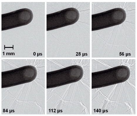
Fig. 50: Time series recording crack propagation in a glass plate initiated by an accelerated bolt. The pictures were acquired via single-bunch imaging at beamline ID19 (from [3]). |
Going to ultra-fast imaging is another major objective for the coming years, in particular within Phase II of the upgrade programme. Feasibility tests at ID15 and ID19 have already demonstrated that the pulsed time-structure of the ESRF’s synchrotron light can be used to capture snapshot images of ultra-fast processes such as crack propagation, shown in Figure 50. Using the isolated X-ray flash from a single-bunch of electrons in the storage ring, which is as short as 140 ps, allows access to a completly new time domain for hard X-ray imaging [3].
M. Cotte
References
[1] A. Horng, E. Brun, A. Mittone, S. Gasilov, L. Weber, T. Geith, S. Adam-Neumair, S.D. Auweter, A. Bravin, M.F. Reiser and P. Coan, Cartilage and soft tissue imaging using X-rays: propagation-based phase-contrast computed tomography of the human knee in comparison with clinical imaging techniques and histology, Investigative Radiology 49, 627-34 (2014).
[2] B. Laforce et al., Nanoscopic X-ray fluorescence imaging of meteoritic particles and diamond inclusions, Analytical Chemistry 86, 12369–12374 (2014).
[3] A. Rack et al., Exploiting coherence for realtime studies by single-bunch imaging, Journal of Synchrotron Radiation 21, 815-818 (2014).
partners


European Synchrotron Radiation Facility - 71, avenue des Martyrs, CS 40220, 38043 Grenoble Cedex 9, France.