- Home
- News
- Spotlight on Science
- Localised deformation...
Localised deformation in nickel-titanium shape memory alloy wires
03-08-2016
Super elastic nickel-titanium (NiTi) alloys can tolerate large deformations due to a stress-induced martensitic transformation. When NiTi wire is stretched, a propagating macroscopic transformation front moves along the wire, separating transformed and untransformed regions, unlike other materials which deform homogenously. 3D-XRD stress tomography provided scientists with stress and strain tensors in austenite grains near this propagating front, and they could reconstruct a complete 3D picture of the front using digital image correlation and finite element modelling.
From eyeglass frames to jet engines, shape memory alloys are used in a wide range of applications because of their unique mechanical properties. Nickel-titanium (Nitinol) wires can withstand stretching by almost 10% and then come back to their original length upon unloading or heating. After many repeated cycles of stretching, fatigue sets in and wires can eventually wear out as cracks appear before they fail. To design materials with even better performance, a research team from Prague set out to determine what happens inside the tensioned wire while the stress driven martensitic transformation takes place. A joint PhD project was set up between the ESRF, the Institute of Physics CAS and the Czech Technical University in Prague to investigate shape memory alloys using X-ray diffraction methods.
The propagation of martensite band fronts in NiTi has previously been studied using a wide range of experimental techniques [1] and represents a challenge for mechanical modelling. In flat sheet samples, the interface between the deformed and undeformed material is sharp and makes an angle of about 55° to the loading direction. For wires and bars, the interface appears to be broad and perpendicular to the loading direction. High-energy synchrotron X-rays are an ideal tool to look inside these wires during loading tests [2].
 |
|
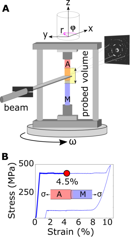
Figure 1: A) The 3D-XRD experiment, and B) the stress strain curve. |
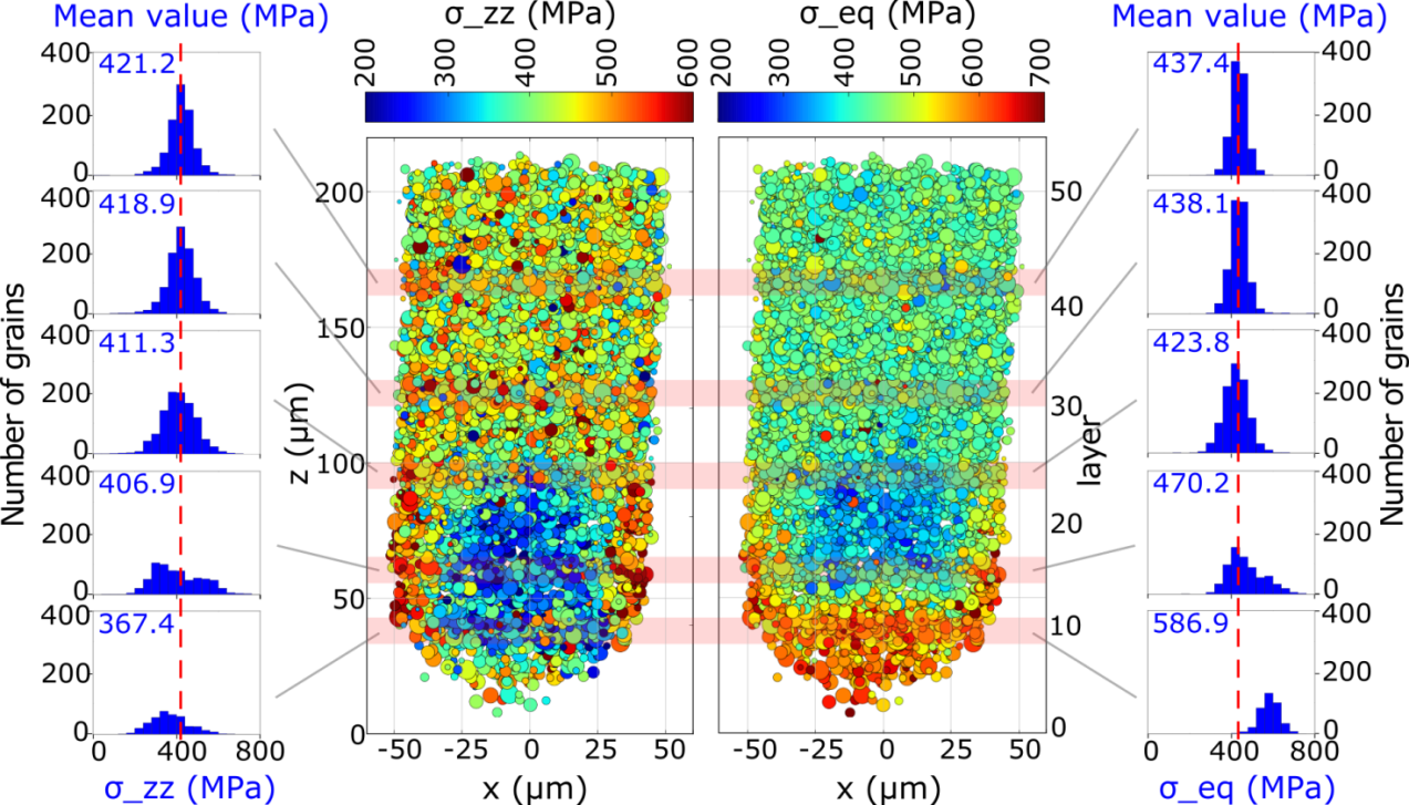
X-ray diffraction can be used to detect the mechanical stress and whether the sample has gone through the martensitic transformation. 3D-XRD [3], which has been developed at the ESRF, is based on single crystal diffraction and gives access to the full stress tensor, position, and orientation of each polycrystal grain in the sample. Employing 3D-XRD at beamline ID11, the team had to measure individual diffraction spots from the grains and then index these spots in order to determine the crystal lattice of each of the approximately 15,000 grains (figure 1). This was only possible for the grains on the undeformed side of the propagating front: after the martensitic transformation many different orientations of the monoclinic martensite phase appear inside a single parent grain and this causes the diffraction spots to be smeared out. Figure 2 shows the key result of the 3D-XRD experiment: a strong internal stress gradient and elevated shear stresses are seen at the edge of the nose-cone shaped macroscopic interface which forms the core of the propagating martensite band front.
 |
|
Figure 2: The 3D-XRD results: stress in grains as a function of position in the wire near the interface. On the left σzz is the stress in the loading direction and on the right σeq is the Von Mises equivalent stress which drives the martensitic transformation. Histograms show the distribution of stresses within the marked horizontal sections through the wire. |
These 3D-XRD results gave a tantalising glimpse of what is happening in the sample, but only the part of the sample which has not transformed is visible in the picture. In this experiment, the grain size of the sample was tailored for the 3D-XRD experiments by a rapid joule heating process. The grains were large enough to be resolved but small enough to provide high resolution pixelation of the 3D volume to map out the full tensorial stress state, but only on the undeformed side of the interface.
 |
|
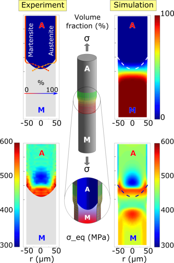
Figure 3: Martensite band front propagating in a NiTi wire. Left: phase fractions (above) derived from powder diffraction and Von Mises equivalent stresses (below) from 3D-XRD. Right: finite element simulation results for the same quantities. |
To elucidate what was happening in the martensitic part of the sample, the team measured surface strains by digital image correlation and they carried out finite element simulations using an original shape-memory alloy model [4]. To compare the simulations with experiment (Figure 3), the researchers faced a new problem: 3D-XRD was giving the stress in each individual grain but the simulation computes a continuous field that does not account for the local granular orientations. This is a common problem in engineering materials research: large-scale components are usually modelled as a continuum instead of an ensemble of polycrystal grains. The macroscopic internal stress field in the wire was obtained by averaging and interpolation of the local stresses in the grains. The very large number of small grains which were measured in this experiment was essential to retrieve a reliable value for the average. In the end, the agreement was excellent and confirms that finite element modelling is able to capture the key features of the super elastic deformation, which is localised in macroscopic transformation fronts in tensioned NiTi wires.
Principal publication and authors
Grain-resolved analysis of localized deformation in nickel-titanium wire under tensile load, P. Sedmák (a,c), J. Pilch (a), L.Heller (a), J. Kopeček (a), J. Wright (b), P. Sedlák (d), M. Frost (d), P. Šittner (a), Science 353, 559-562 (2016); doi: 10.1126/science.aad6700.
(a) Institute of Physics of the Czech Academy of Sciences, Prague (Czech Republic)
(b) ESRF
(c) FNSPE, CTU Prague (Czech Republic)
(d) Institute of Thermomechanics of the Czech Academy of Sciences, Prague (Czech Republic)
References
[1] C.B. Churchill, J.A. Shaw, M.A. Iadicola, Experimental Techniques 33, 70-78 (2009).
[2] P. Sedmák, P. Šittner, J. Pilch, C. Curfs, Acta Mater. 94, 257-270 (2015).
[3] H.F. Poulsen, Three-dimensional X-ray diffraction microscopy: mapping polycrystals and their dynamics, Springer, Berlin (2004).
[4] P. Sedlák, M. Frost, B. Benešová, P. Šittner, T. Ben Zineb, Int. J. Plast. 39, 132–151 (2012).
Related content
- The complete dataset obtained from the 3DXRD is available online together with further information: http://ofm.fzu.cz/localized%20deformation%20of%20NiTi%20in%20tension
Top image: Visualisation of grain-resolved internal stress in a nickel-titanium alloy undergoing deformation through 3D-XRD.
partners


European Synchrotron Radiation Facility - 71, avenue des Martyrs, CS 40220, 38043 Grenoble Cedex 9, France.