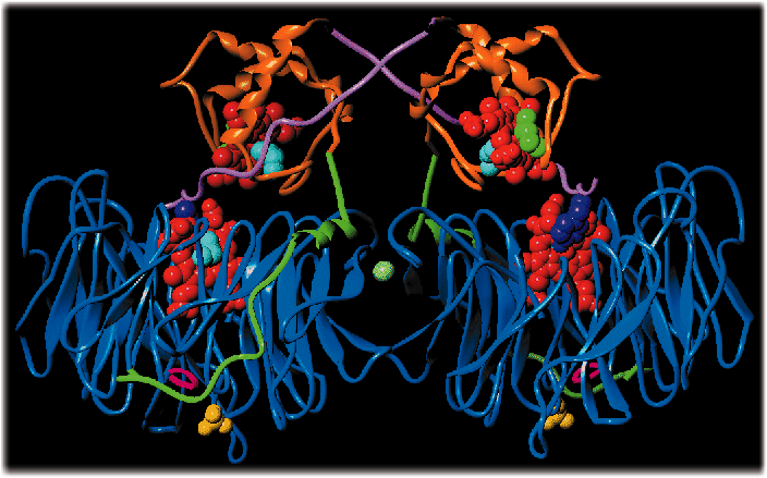
Figure 19
Fig. 19: Ribbon representation of the secondary structure of the C tracing of nitrite reductase from Pseudomonas aeruginosa. The c and d1 hemes are represented in red, the c-domain in orange, the d1-domain in blue and the c-d1 linker in green.
| back to: Seeing an Enzyme at Work: Conformational Changes Occurring Upon Nitrite Reduction |
partners


European Synchrotron Radiation Facility - 71, avenue des Martyrs, CS 40220, 38043 Grenoble Cedex 9, France.