- Home
- Users & Science
- Scientific Documentation
- ESRF Highlights
- ESRF Highlights 2017
- Enabling technologies
- Smallest ever nanofocus for high-energy nanoimaging using X-ray mirrors
Smallest ever nanofocus for high-energy nanoimaging using X-ray mirrors
A sub-13 nm diffraction-limited X-ray focus size with six billion photons per second was reached at the ID16A nanoimaging beamline using a high precision, graded multilayer Kirkpatrick-Baez mirror system. This was made possible by significant advances in mirror fabrication technology along with a highly optimised and stable beamline optical system.
The availability of high-energy X-ray nanobeams for routine use offers new opportunities across a wide range of scientific fields. However, forming nanobeams with suitable photon flux at high-energies is an engineering challenge. As part of the newly commissioned 185 m-long ID16A nanoimaging beamline at the ESRF, a new optical system consisting of a pair of elliptically figured Kirkpatrick-Baez (KB) mirrors (Figure 146) operating at 33.6 keV delivered a sub-13 nm high-energy X-ray focus. An impressively high flux of six billion photons per second was efficiently concentrated in the focus (Figure 147). This is the smallest and brightest focus ever reported for energies above 20 keV.
 |
|
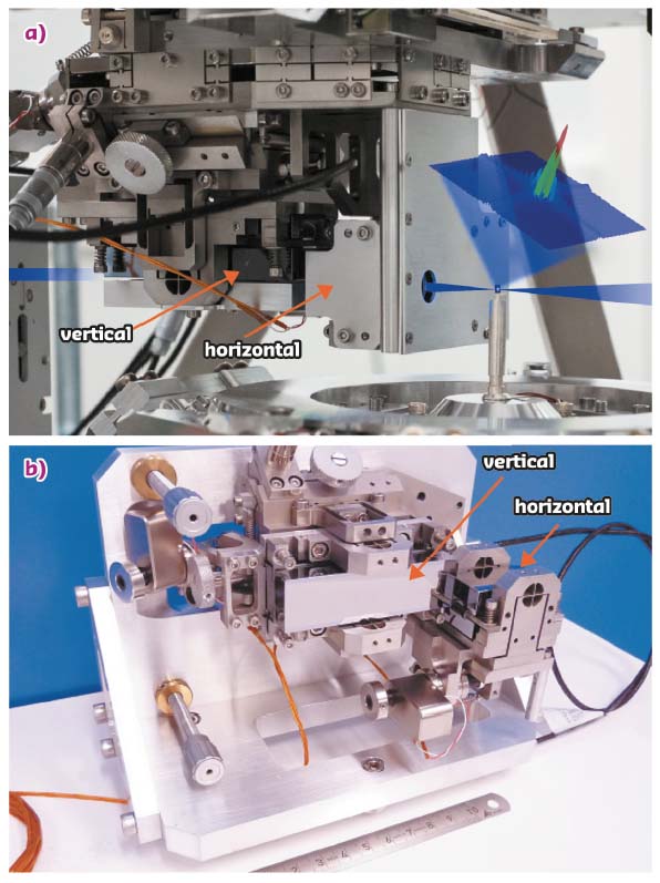
Fig. 146: The KB system on ID16A. a) Complete set-up with visualisation of the X-ray beam path (blue) and the reconstructed focal spot image. b) View of the KB mechanics including the horizontal and vertical mirrors. |
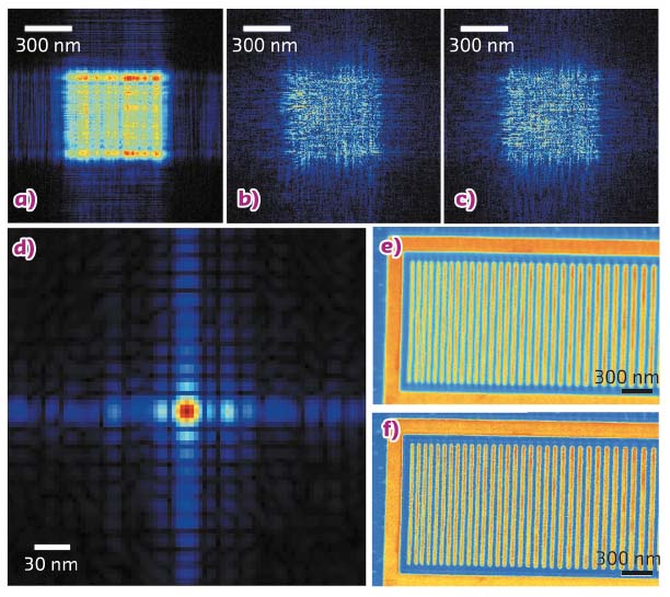
This diffraction-limited focus was carefully characterised not only by sharp-edge X-ray fluorescence scans but also by ptychography measurements. Compared to the edge scan measurements, which tend to overestimate the focal spot size, ptychography provides a more precise evaluation of the wavefront. Thus, ptychography was used to retrieve the wavefront at the sample position, taking into consideration partial coherence effects of the beam, as shown in Figures 147a-c. The measurements were taken in a defocused position and the retrieved wavefront was numerically propagated back to the focus position by Fresnel propagation. To confirm the retrieved focus (Figure 147d), an X-ray fluorescence imaging experiment was performed on a lithographic sample in the focus position (Figure 147e). Figure 147f shows the deconvolved image using the focus determination from the ptychography measurements and shows further improvement on the image resolution (Figure 147).
 |
|
Fig. 147: The X-ray wavefront. The coherent modes of the beam retrieved by ptychography at the sample position: a) main mode (87% power), b) secondary (7% power) and c) tertiary modes (6% power). d) The retrieved focus. e) Original and f) deconvolved XRF images. |
Figure 146a shows the KB system used for X-ray focusing at the ID16A beamline, consisting of two mirrors: one horizontally deflecting and one vertically deflecting (Figure 146b). The KB system required mirrors with unprecedented surface perfection. The horizontal mirror is 36 mm long while the vertical one is 70 mm long [1]. The elliptical cylinder figured substrates were prepared by a commercial supplier, using polishing techniques based on technologies developed at Osaka University. The mirrors were coated at the ESRF’s Multilayer Laboratory with 120 periods of W/B4C, with a d-spacing of 2 nm. The main challenge was to coat both mirrors with a steep thickness gradient while using a 7 m-long deposition machine. The coating increases the useful numerical aperture of the optics, which reduces the diffraction-limited focus size produced by the mirrors. An in-house-designed mechanical assembly was developed to support and align the mirrors.
The ID16A nanoimaging beamline was built as part of the ESRF Phase I Upgrade. The beamline features a precisely temperature-controlled environment and vibrational stability, which makes high-resolution imaging possible. A cryogenic setup is also available for radiation sensitive samples. The available techniques include full-field phase-contrast imaging, X-ray fluorescence imaging, and ptychography, all being combined with tomography to obtain 3D imaging. Applications include the study of nanotechnological materials, environmental and biological samples [2].
X-ray imaging techniques, where resolution depends on the focus size, can fully benefit from the diffraction-limited nanobeam available at the ID16A beamline. The high energy and high flux available in such a tiny beam allows imaging of thick or highly absorbing samples with high spatial resolution, in addition to extending the range of detectable elements by X-ray fluorescence imaging [2]. The spatial resolution currently achievable at such a beamline offers the possibility of carrying out X-ray imaging experiments with resolution higher than that provided by super-resolution optical microscopy, without the requirement of any fluorescence markers. The present achievement is relevant not only for the X-ray optics and X-ray imaging communities, but also for many other scientific fields, which can benefit from probing samples with unprecedented precision.
Principal publication and authors
Efficient concentration of high-energy X-rays for diffraction-limited imaging resolution,
J. C. da Silva (a), A. Pacureanu (a), Y. Yang (a), S. Bohic (a, b), C. Morawe (a), R. Barrett (a) and P. Cloetens (a), Optica 4(5), 492-495 (2017); doi: 10.1364/OPTICA.4.000492.
(a) ESRF
(b) EA-7442 RSRM, Université Grenoble Alpes, Grenoble (France)
References
[1] C. Morawe et al., Proc. SPIE 9588, 958803 (2015).
[2] J. C. da Silva et al., Proc. SPIE 10389, 103890F (2017).
partners


European Synchrotron Radiation Facility - 71, avenue des Martyrs, CS 40220, 38043 Grenoble Cedex 9, France.