- Home
- Users & Science
- Scientific Documentation
- ESRF Highlights
- ESRF Highlights 2013
- Enabling technologies
- New KHz frame-rate detector applied to time-resolved EXAFS
New KHz frame-rate detector applied to time-resolved EXAFS
We have developed a new high frame rate detector based on the FReLoN design and featuring a linear image sensor coupled to an electronic shutter. This new detector has been developed specifically for fast time-resolved energy dispersive EXAFS at beamline ID24 [1].
ID24 is an energy-dispersive X-ray absorption beamline (XAS) that has recently been rebuilt through the ESRF Upgrade Programme. It offers the user community new opportunities for investigating matter at extreme conditions of pressure, temperature and magnetic field. These experiments combine very stringent time resolution (usually below the millisecond) coupled to a small focal spot (few micrometres). Detector readout speed is the limiting factor for time resolution.
The new EXAFS detector is based on the layout of the previous model, with a phosphor screen X-ray converter coupled to a CCD camera by a tandem lens system. The optical front-end was totally redesigned in order to fit the horizontal beamsize that increases from 50 mm to 100 mm and now includes a custom high-resolution front lens. The lens assembly was designed so as to enable spatial resolutions up to 4 k pixels, if needed. The system includes a motorised focusing and a motorised iris diaphragm for fine resolution and sensitivity adjustments.
The workhorse detector during the past 10 years has been the ESRF FReLoN camera, equipped with a conventional 2D CCD area image sensor used as a linear image sensor via line binning [2]. For the upgraded ID24, the number of photons transmitted by the sample is close to ~ 1014 ph/s. However, the 1 KHz acquisition frequency was a limitation of the present FReLoN camera [2], which did not allow efficient detection for such a high photon count. To go beyond the KHz frame-rate while maintaining a high dynamic range, linearity and spatial (or equivalently, energy) resolution, a new system had to be designed that would allow single images to be recorded without lag or aliasing of images. These features were considered essential for sub-ms EXAFS.
 |
|
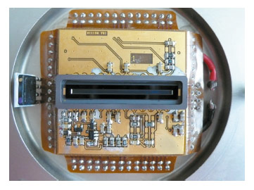
Fig. 151: The S11156 Hamamatsu sensor integrated in the FReLoN camera. |
A new camera was developed, based on the linear CCD image sensor 11156 from Hamamatsu, consisting of an array of 2048 pixels of dimensions 14 x 1000 µm2 (Figure 151). The S11156-2048 chip utilises a resistive gate structure that allows high-speed transfer, with “on chip” electronic shutter function, offering significantly reduced image lag (less than 0.1%). Its minimum exposure time is 30 μs, and it has a true dynamic range of 14 bit. With its back-thinned structure, this CCD also offers high sensitivity (> 80% quantum efficiency) from the UV to the NIR region of the spectrum. Figure 152 illustrates the quality of data collected on pure Cu foil on ID24.
 |
|
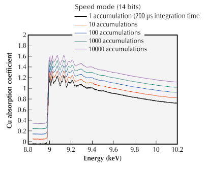
Fig. 152: Cu K –edge EXAFS recorded at ID24 by averaging an increasing number of accumulations of 200 μs each. The EXAFS features are already clearly visible in the single shot spectrum (black). |
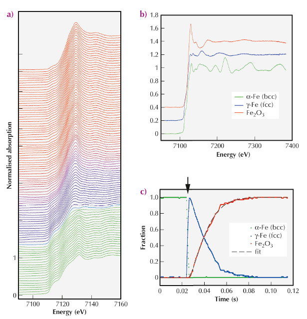
To demonstrate the potential of the new “Hamamatsu sensor” FReLoN camera, a time-resolved XAS study of iron oxidation at high temperature was performed at beamline ID24. A pure Fe foil of 5 μm thickness exposed to normal atmosphere (oxygen fugacity = –0.7) was heated with a highly defocused 5 W IR laser. A polychromatic X-ray beam was focused down to about 5 x 4 μm2 on the sample in order to minimise probed thermal gradients. The foils temperature was raised from room temperature to about 1200 K within a few ms. Figure 153a shows a sequence of Fe K-edge (7.1 keV) XAS spectra, recorded every 200 µs allowing the kinetics of the chemical reaction to be followed in situ. Spectral features change as the reaction evolves from ambient temperature (data in green) to the highest temperatures (data in red). The EXAFS spectra were processed with a standard linear combination fit technique to determine the relative fractions of the principal components (Figure 153b) – namely, α-Fe (body-centred cubic structure), γ-Fe (face-centred cubic polymorph of iron) and Fe2O3 hematite. The evolution with time of the relative fractions of the three phases is shown in Figure 153c.
 |
|
Fig. 153: a) Sequence of Fe K-edge XAS, acquired every 200 μs. b) Principal components of the dataset: α-Fe, γ-Fe and Fe2O3. c) Time evolution of relative fractions. The black arrow indicates the onset of laser heating. Courtesy of I. Kantor. |
The performance of the new detector appears to be very attractive for a number of applications beyond EXAFS, for example, for computed tomography imaging of fast-moving samples that are particularly important for in vivo physiological studies.
Authors
J.-C. Labiche, E. Collet, L. Siron, J.J. Thevenin, C Ponchut, A. Homs, M.C. Dominguez, J. Borrel, C. Jarnias, C. Strohm, I. Kantor, O. Mathon and S. Pascarelli.
ESRF
References
[1] S. Pascarelli et al., J. Synch. Rad. 13, 351 (2006).
[2] J.C. Labiche et al., Rev. Sci. Instrum. 78, 091301 (2007).
partners


European Synchrotron Radiation Facility - 71, avenue des Martyrs, CS 40220, 38043 Grenoble Cedex 9, France.