- Home
- Users & Science
- Scientific Documentation
- ESRF Highlights
- ESRF Highlights 2000
- Microfocussing and Imaging
- In situ Real-time Radiography for Process Optimisation Foaming of Metal
In situ Real-time Radiography for Process Optimisation Foaming of Metal
It has been shown that in situ real-time radiography is extremely useful for the observation of the foaming process in liquid metals. In addition, ex situ micrometre computer tomography on pre-foamed samples provides complementary information about the 3D structure in early foaming stages.
Solid metal foams have attracted much attention because of their potential applications, e.g. in automotive and aerospace industries, where their specific structure makes them useful for lightweight construction or crash energy absorption. Several ways of producing metal foams are available. The method developed at Fraunhofer-IFAM consists of mixing a metal powder (pure elements such as Zn or alloys such as AlSi7 and Al6061) with a gas releasing foaming agent [1]. The resulting powder blend is compacted by uniaxial hot pressing into small tablets. It is possible to join two faces of this material to face sheets during the compaction process by roll-cladding. The resulting sandwich structures are of special technological interest since they allow the encapsulation of the foam structure under a closed surface and also show good mechanical properties.
Although metal foams are rapidly becoming industrially important, surprisingly little is known about the physical processes governing their formation. In order to improve further such foams their liquid counterpart has to be investigated thoroughly, which is difficult to perform owing to the opacity, reactivity, high temperature and low electrical resistivity of metal melts. Therefore, observation methods applicable for studying aqueous foams such as resistance, fluorescence or light scattering measurements are unsuitable. In contrast, the use of synchrotron radiography allows an unprecedented insight into evolving metal foams.
For heating the samples, a high-temperature furnace was specially designed to permit the X-ray beam transmission. The photon beam was monochromatised to 33 keV and used to generate an absorption radiograph of the samples on the electronic detector system (a scintillator screen coupled to a FReLoN-camera by light optics). The effective pixel size was 40 µm and the whole CCD camera array was read out at frequencies between 2 and 3 Hz.
Foaming of the samples was triggered in the furnace by heating the tablets to the melting range of the alloy. This caused partial melting of the alloy, hydrogen release by the foaming agent and the formation and inflation of bubbles with a corresponding volume expansion of a factor 5 within a period of a few minutes.
Figure 112 shows some stages of the foaming process of an AlSi7 sandwich structure [2]. An AlSi7 core is joined to two face sheets of an alloy of a higher melting point. Because of the high temperature gradient, the core first melts near the conductive sheets (a), thus it starts to foam there first. After this, the entire core layer foams (b)-(c).
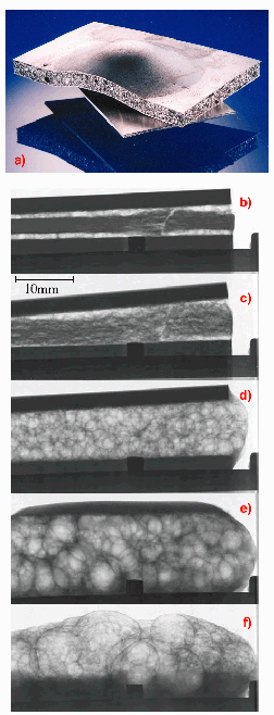 |
Fig. 112: Foaming of an aluminium foam sandwich
(a) Al foam sandwich structures for light-weight applications (b)-(f) real-time radiography with images of different stages of the development of fissures and pores, of melting of the covering sheet areas and drainage up to the collapse of the foam. |
In contrast to other metals like zinc, the release of the hydrogen takes place before the melting point of the alloy is reached. Therefore, during the first phase the hydrogen causes the appearance of fissures in the material. Only in a later stage, after the development of a liquid phase, a network of bubbles forms which subsequently shows effects of drainage and partial collapse of the bubble structure during further evolution, as shown in Figure 112 (e)-(f).
From the series of radiographic images, phenomena such as bubble creation and growth, rupture of films, local topological rearrangements, collapse and drainage of the foam can be studied. Furthermore, the great wealth of detail in the images allows conclusions to be drawn about the stabilisation and destabilisation mechanisms governing the foaming process. A more exhaustive discussion is given in reference [3].
Figure 113 shows rendered 3D-images of reconstructed volumes of various aluminium foam samples at different stages of the foaming process. The titanium-containing foaming agent residues are strongly absorbing, and indicated by the red regions. At the beginning of the foaming process, the developing pores have the shape of micro-fissures (a). Upon development of the liquid phase, the pore morphology is transformed into bubbles (d).
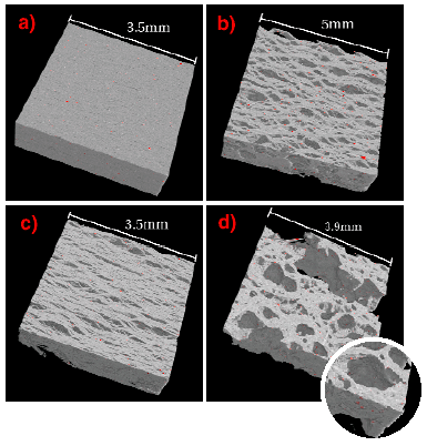 |
Fig. 113: Rendered 3D images of the CT-reconstructed volumes of Al foam samples corresponding to different stages of the foaming: relative density (a) 99 %, (b) 70 %, (c) 56 % and (d) 44 % of the volume material. The regions highlighted in red are strongly absorbing, titanium-containing residues of the foaming agent.
|
The foaming behaviour could be observed in situ and in real time by help of this kind of analysis. The method provides new knowledge, regarding the effect of the blowing gas hydrogen on the foaming process, the dynamics of pore formation, the size-related pore distribution and provides opportunities for optimisation of the foaming processes. This will be the objective of further radiographic analysis, in particular with regard to the temperature-time behaviour of the features of the structure and the material.
References
[1] J. Banhart, Europhysics News 30, 17 (1999).
[2] J. Banhart, H. Stanzick, L. Helfen and T. Baumbach, Advanced Engineering Materials, submitted for publication.
[3] J. Banhart, H. Stanzick, L. Helfen and T. Baumbach, Applied Physics Letters 78 (8), (2001).
Principal Publication and Authors
L. Helfen (a,b), H. Stanzick (c), J. Banhart (c) and T. Baumbach (d).
(a) Fraunhofer-Institute for Non-destructive Testing, Saarbrücken (Germany)
(b) ESRF
(c) Fraunhofer-Institute for Advanced Materials, Bremen (Germany)
(d) Fraunhofer-Institute for Non-destructive Testing, Dresden (Germany)
partners


European Synchrotron Radiation Facility - 71, avenue des Martyrs, CS 40220, 38043 Grenoble Cedex 9, France.