Microscope Options
The sample positioning is recorded by a long working distance zoom microscope, that presently watches the interaction point from above and is equipped with a colour CCD camera and frame grabbing system.This is replaced by a new system to allow more flexibility with the positioning of the microscope. The new microscope can view the sample from the same direction as the x-ray beam with a variable angle vertically. It uses a fixed magnification with several fields-of-view to choose.
 view of the sample position (171 kB)
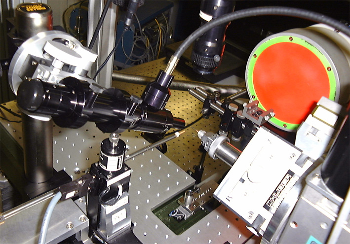
In the above picture one can see the x-ray beam pipe coming from left below and hitting the sample on a small goniometer head. Above the new microscope is positioned to look on the sample with an angle of 25 to 70 degrees. On the right hand side the lately installed Eulerian cradle is to be seen.
characteristics of the zoom microscope:
characteristics and improvement of the new Infinity(c) microscope
The philosophy is to choose the magnification, working distance or illiumination type at the beginning of the experiment and then align the center of the diffractometer and not change the setup during the run. The problem with the abolished zoom feature can be overcome by prealigning the sample on the ex-situ microscope with goniometer holder. In that way much time is saved and sample changes are simplified.
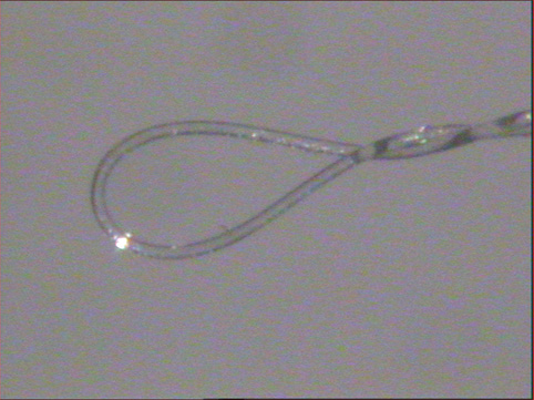
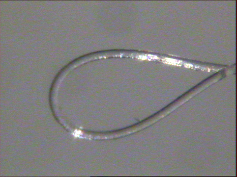
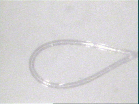
The following series of pictures show the image from a crystal loop positioned at the sample position together with the performance figures of the system
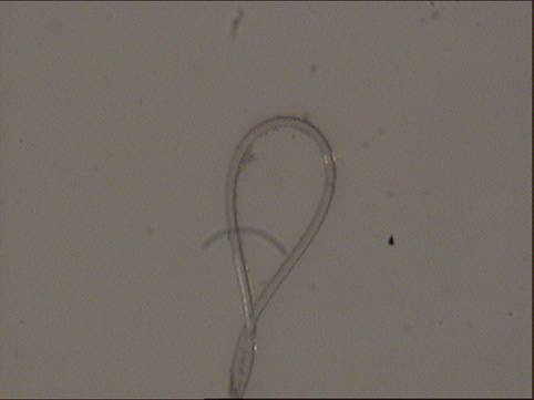 |
old microscope
|
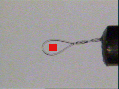 |
new microscope
|
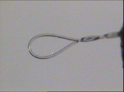 |
LWD 3 objective
|
 |
LWD 2 objective + 2x extension
|
 |
LWD 3 objective + 2x extension
|
A coaxial illumination option is available, which avoids the difficulties of positioning the light fiber around the close packed sample area. The disadvantage is a picture with less contrast due to the flat reflective illumination and some stray scatter in the microscope tube.
 |
LWD 3 objective + 2x extension
|
-
- fixed position vertically above the sample
- 4x zoom with smallest field of view about 1.4 x 1 mm
- working distance 90 mm
- heavy mount
- micrometer screw adjustment
- Sony camera 1/2 '' colour
- variable position in a plane with the incoming x-ray beam (25 to 70 degrees)
- fixed focus, different magnification options (currently a 2x objective LWD2, 3x objective LWD3 and 2x extension tube)
- working distance 43 mm (LWD3) or 69 mm (LWD3)
- possible coaxial illumination
- compact mount and stable fixation on a StableRod(c) system
- Watec camera 1/3 '' colour
partners


European Synchrotron Radiation Facility - 71, avenue des Martyrs, CS 40220, 38043 Grenoble Cedex 9, France.