- Home
- Users & Science
- Scientific Documentation
- ESRF Highlights
- ESRF Highlights 2002
- Soft Condensed Matter
- Twisted Plywood Pattern of Collagen Fibrils in Fish Scales
Twisted Plywood Pattern of Collagen Fibrils in Fish Scales
The mineralised collagen fibril represents the building unit of several calcified tissues, such as tooth, bone and calcified tendons. Different patterns of fibril arrays have been described [1]. The results of a number of morphological and structural investigations performed on various mineralised tissues have indicated a close structural relationship between the nanocrystalline carbonated apatite and collagen molecular packing as collagen acts as a template for the deposition of the mineral phase [1,2]. We have shown that the local distribution and orientation of collagen fibrils and apatite crystals in mineralised tissues can be successfully investigated by means of micro-small-angle X-ray scattering (µSAXS) [3]. Here we describe the use of µSAXS on ID13 to study the distribution and orientation of collagen and apatite in fish scales.
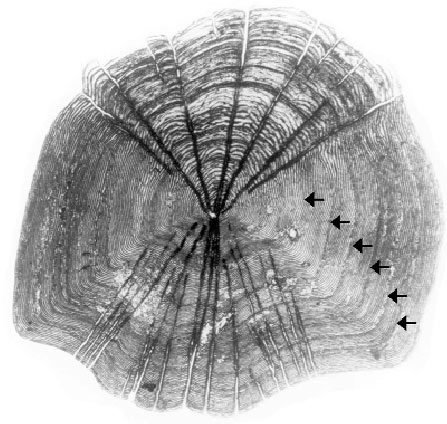
A typical scale from a teleost fish (Leuciscus cephalus) is shown in Figure 25. These scales grow and are mineralised continuously throughout life. The annuli, some of which are labelled with arrows in the figure, mark the pauses of growth in winter. µSAXS patterns with a 7 µm beam were obtained from a single scale by step-scanning over areas as large as 600 x 600 µm. After every step a 2D-µSAXS pattern was obtained with a CCD detector.
 |
|
Fig. 25: Optical micrograph of a typical scale from Leuciscus cephalus. The annuli are indicated by arrows. |
The µSAXS patterns from most of the examined areas exhibit a distribution of intensity of the collagen reflections according to five directions of preferential orientation, at 36° from each other (Figure 26). The data suggest that this pattern is due to a plywood arrangement of collagen fibrils in successive layers parallel to the surface of the scale. The fibrils are strictly aligned in each layer and the alignment rotates by 36° in successive layers, according to a discontinuous twist that generates a symmetric plywood pattern. Evidence for a preferred alignment of c-axes of the hexagonal apatite crystallites parallel to the collagen fibrils was obtained from the 002 reflection. These are the very first diffraction data, which unambiguously confirm the presence of these peculiar structures, and suggest that this kind of investigation represents a powerful tool to study plywood arrangements in biological tissues.
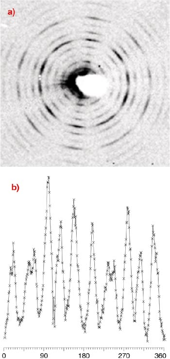 |
Fig. 26: Micro X-ray small-angle scattering pattern recorded from a decalcified scale. The reflections correspond to the orders of collagen (periodicity: 64 nm). The reflections are preferentially oriented along 5 preferential directions. The relative integrated intensity distribution along the Debye ring of the sixth order collagen reflection is reported in b) as a function of azimuthal angle. |
References
[1] S. Weiner and H.D. Wagner Annu. Rev. Mater. Sci. 28, 271 (1998).
[2] A. Ascenzi, A. Benvenuti, A. Bigi, E. Foresti, M.H.J. Koch, F. Mango, A. Ripamonti, and N. Roveri, Calcif. Tissue Int. 62, 266-273 (1998).
[3] A. Bigi, M. Burghammer, R. Falconi, M.H.J. Koch, S. Panzavolta and C. Riekel, J. of Struct. Biol. 136, 137 (2001).
Authors
A. Bigi (a), S. Pannzavolta (a), M. Burghammer (b), C. Riekel (b), R. Falconi (a), M.H.J. Koch (c).
(a) University of Bologna (Italy)
(b) ESRF
(c) EMBL, Hamburg Outstation, Hamburg (Germany)
partners


European Synchrotron Radiation Facility - 71, avenue des Martyrs, CS 40220, 38043 Grenoble Cedex 9, France.