Figure 91
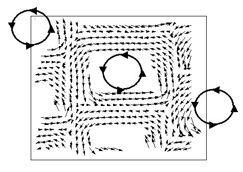
Fig. 91: Map of local cellulose fibril orientation in single wood cell. Following the arrows readily yields the trace of the fibrils around the cell wall.
| back to: Imaging of the Helical Arrangement of Cellulose Fibrils in Wood Cells |
partners


European Synchrotron Radiation Facility - 71, avenue des Martyrs, CS 40220, 38043 Grenoble Cedex 9, France.