Figure 90
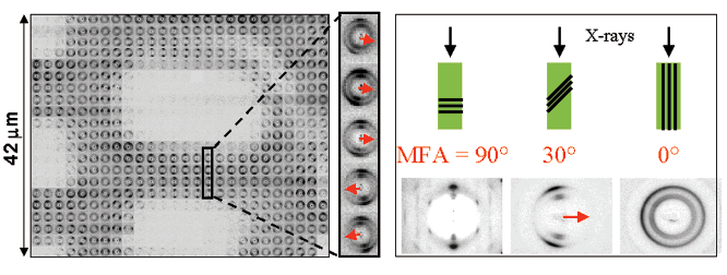
Fig. 90: Mesh scan of a single Picea abies wood cell on a 2x2 µm2 grid. A single line of patterns has been enlarged. A red arrow symbolises the orientation of the microfibrils relative to the cell wall. The determination of this orientation, as defined by the MFA, is shown schematically on the right. A perfect powder pattern is observed when the microfibrils (black lines) are parallel to the incoming X-ray beam (MFA = 0°) while a fibre diffraction pattern is observed when they are at right angles (MFA = 90°). The distortion allows a determination of the MFA.
| back to: Imaging of the Helical Arrangement of Cellulose Fibrils in Wood Cells |
partners


European Synchrotron Radiation Facility - 71, avenue des Martyrs, CS 40220, 38043 Grenoble Cedex 9, France.