Figure 30
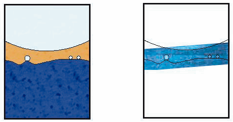
Fig. 30: Liquid-liquid interface as observed using the X-ray camera. This figure denotes a snapshot of the phase separation in dilute (blue) and concentrated (gold) phases solutions. For clarity, the coloured schematic (left) highlights the two phase region observed in the photograph opposite. The upper meniscus, liquid-liquid interface, and ammonia bubbles forming at the interface are shown. The width of the sample container is 1 cm.
| back to: X-ray Absorption Spectroscopy Investigations of Metal-ammonia Solutions |
partners


European Synchrotron Radiation Facility - 71, avenue des Martyrs, CS 40220, 38043 Grenoble Cedex 9, France.