- Home
- News
- General News
- Synthesizing the...
Synthesizing the most natural of all skin creams
17-03-2009
Research done at the ESRF by scientists from Leiden University could help millions of people with skin problems
Even after nine months soaking in the womb, a newborn’s skin is smooth – unlike an adult’s in the bath. While occupying a watery, warm environment, the newborn manages to develop a skin fully equipped to protect it in a cold, dry and bacteria-infected world. A protective cream called Vernix caseosa (VC), which covers the fetus and the newborn, aids in the growth of skin both before and after birth. VC provides ‘waterproofing’ in utero, allowing skin to grow in wet conditions, while after birth it hydrates and cleanses, even healing when applied to ulcers. Prof. Joke Bouwstra, a specialist in the skin barrier and its synthesis at Leiden University, and her colleague Robert Rißmann set out to study VC in detail and has produced a synthetic version of this natural buttery ointment which shows the same structure and unique properties. As well as helping pre-term babies develop essential protection against temperature changes, dehydration and infection, artificial VC could also benefit sufferers of skin disease.
Like most moisturising creams, VC is mostly water. Its outstanding properties come from the addition of just 10% each of lipid molecules and dead skin cells (corneocytes), so the exact composition of the mixture is important.
For the lipids, X-ray diffraction measurements at the Dutch/Flemish DUBBLE beamline at the ESRF (European Synchrotron Radiation Facility) allowed the Leiden researchers to find the proportions of the various forms in the cream, even distinguishing between complex molecules differing in chain length.
The corneocytes were also studied using electron microscopy, yielding their size, shape and water content.
But equally important is how the mixture arranges itself. Lipid molecules are shaped something like lollipops, with a round end that prefers to be surrounded by water and a stick which prefers to make a raft with other lollipop sticks. VC contains several different lengths of lipids, which form different arrangements as the temperature changes. The result is that VC fulfils different functions inside and outside the womb, just as butter behaves differently in the oven and on the table. Again, the ESRF’s synchrotron light was used to illuminate the corneocytes and lipids together and look for any clumps or other ordering. Once they knew exactly what VC was made of and how it was arranged, they set about creating a synthetic version.
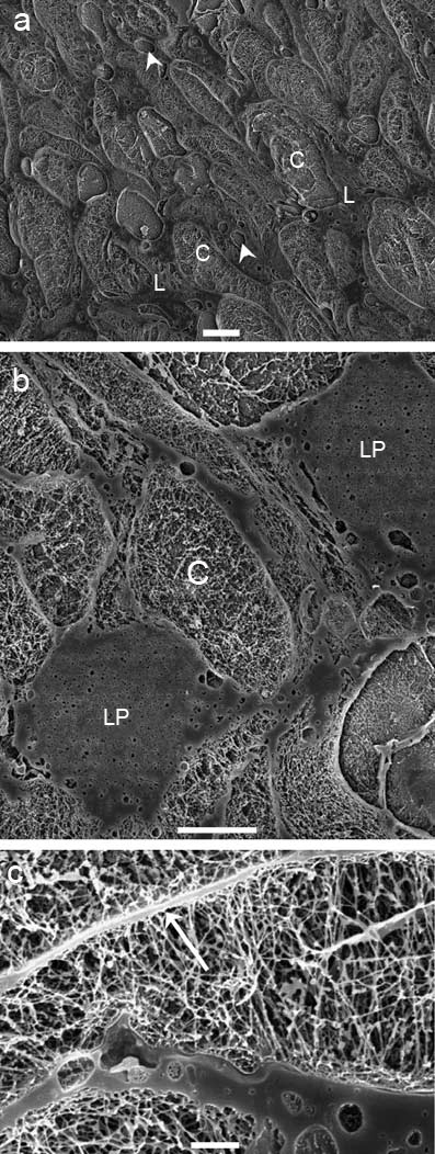 |
Figure 1: Structure of Vernix caseosa (VC) is characterized by corneocytes embedded in lipid domains. Cryo Scanning Electron Microscopy (cryo-SEM) pictures of VC show corneocytes (C), which are surrounded by lipid domains (L). Spherical structures (arrow heads) can also be observed. (a) A certain state of order can be seen. (b) Lipid material is accumulated between corneocytes and form lipid pools (LP) as depicted. (c) underlines the presence of a cornified envelope (arrow) on the boundary of the cell. The inner cellular network consisting of keratin fibers is also clearly visible. Bars = (a) 10 µm, (b) 5mm, and (c) 1 µm. Image courtesy of R.R.Rißmann. |
A readily available natural source of the sort of fat molecules needed is lanolin, the oil found in sheep’s wool, which is currently used as a skin treatment by some nursing mothers. The team isolated the fats which were the closest match to the measurements they had of VC, and used them to create a synthetic solution with the same behaviour. The corneocytes were synthesized by M.H.M. Oudshoorn from the Utrecht University. When combined, these synthetic ingredients made a cream which looked the same using both x-ray measurements and light microscopy as VC, while allowing the researchers to alter the water content and other properties at will. After pre-clinical testing, the developed creams showed great potential for use on disrupted and underdeveloped skin: the skin barrier recovered much more quickly when synthetic VC was applied. These promising results will give rise to future clinical studies, in order to prove the benefits of the newly developed creams in treating healthy, dry and diseased human skin.
 |
Figure 2: Lipids of VC are sandwiched between corneocytes. Micrographs obtained by FFEM depict smooth cell surfaces, indicated by CS. Intercellular lipid material has a very heterogeneous appearance (L). In addition, spherical structures could again be observed (arrow heads). (a) Cell surfaces (CS), characterized by a patchy pattern (arrows), and intercellular lipids (L). The step in the inset of (b) (arrow) indicates a fracture across a lamellar structure, which only occasionally appeared. (c) A high accumulation of lipid (LP) and spherical structures could be observed. Bars = 1 µm. Image courtesy of R.R.Rißmann. |
Reference:
Robert Rißmann, Development of a vernix caseosa substitute. A novel strategy to improve skin barrier function and repair . Ph. D. thesis defended 17 March 2009 at Leiden University, Faculty of mathematics and sciences (The Netherlands).
Thesis supervisors: J. Bouwstra (Leiden University) and W. Hennink (Utrecht University)
Co-supervisor: Ponec (Leiden University)
Images:
Research images
http://www.esrf.fr/Apache_files/comm/dubble/Figure-1.jpg
http://www.esrf.fr/Apache_files/comm/dubble/Figure-2.jpg
Images courtesy of R.R.Rißmann.
The DUBBLE Beamline
http://www.esrf.fr/Apache_files/comm/dubble/AT26686N.jpg
http://www.esrf.fr/Apache_files/comm/dubble/AT26687N.jpg
http://www.esrf.fr/Apache_files/comm/dubble/AT26693N.jpg
http://www.esrf.fr/Apache_files/comm/dubble/AT26689N.jpg
http://www.esrf.fr/Apache_files/comm/dubble/AT30057_8.jpg
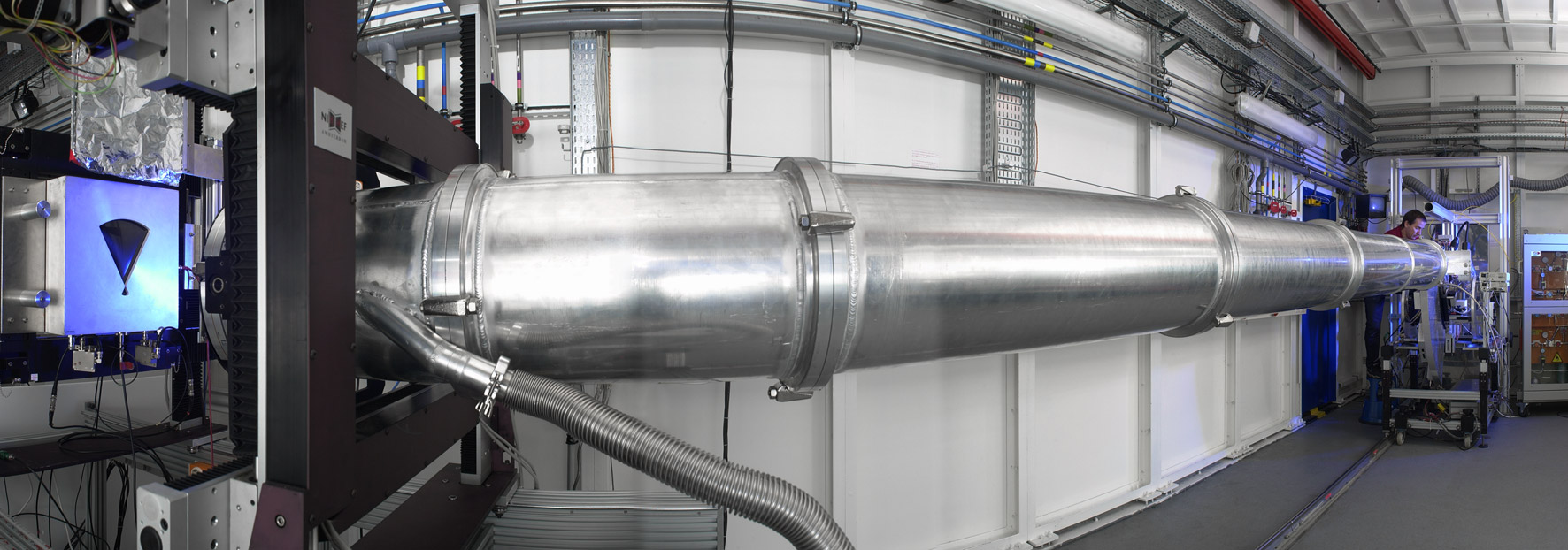
View of the DUBBLE beamlines as they open on to the circular experimental hall. Scientists and technicians use bicycles to get around the experimental hall's 844m circumference.
Images courtesy of Artechnique.
ESRF Facility
http://www.esrf.fr/Apache_files/press/image5.jpg
The ESRF facility from above, showing the main ring where circling electrons generate X-rays, and the surrounding laboratories where this brilliant radiation is used to study atomic structure.
http://www.esrf.fr/Apache_files/comm/dubble/ESRF-main-blue-CMYK.eps
The ESRF logo (vector format)
Images courtesy of ESRF.
University of Leiden
http://www.esrf.fr/Apache_files/comm/dubble/Logo-UL-onder.eps
University of Leiden Logo (vector format)
Image courtesy of the University of Leiden.
For more information, contact:
Montserrat Capellas Espuny, Press Officer, ESRF
e-mail, Tel. +33476 88 26 63
Drs. S. J. Hagers, Press Officer, University of Leiden,
e-mail, Tel. +3171-5274691
Peter van der Wilt, Press Officer, University of Utrecht,
e-mail, Tel. +3130 253 3705
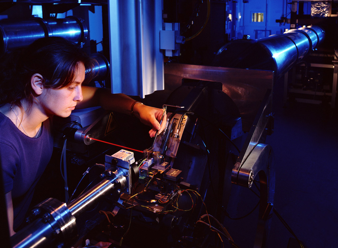
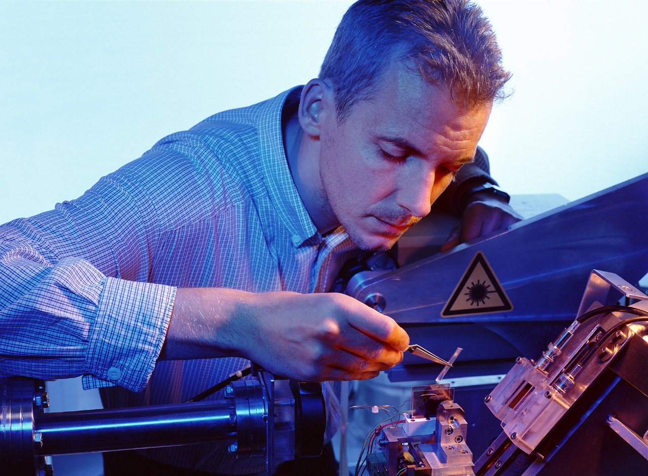
Top image: Scientist working on the DUBBLE beamline
partners


European Synchrotron Radiation Facility - 71, avenue des Martyrs, CS 40220, 38043 Grenoble Cedex 9, France.