- Home
- News
- General News
- #weekendusers: Tracking...
#weekendusers: Tracking nano-precipitates of MRI contrast agents in the liver
16-04-2018
Dr Lorella Pascolo and her all-female team of scientists from Trieste in Italy and Ljubljana in Slovenia have spent 4 days scanning clinical biopsies on the ESRF’s nano-analysis beamline, ID16B. They are trying to determine if there is a relationship between the iron overload and precipitation of widely-used MRI contrast agent gadolinium (Gd) in the liver of oncological child patients.
Gadolinium is used as a contrast agent to improve the visibility of internal organs in magnetic resonance imaging (MRI). Originally considered as innocuous and with rapid clearance on administration, in the last ten years concerns on the safety of gadolinium-based contrast agents (Gd-CAs) have been raised. The first alarm came from the discovery that Nephrogenic Systemic Fibrosis (NSF) was linked to the administration of Gd-CAs. This rare, invalidating and frequently fatal disorder causes fibrosis of the skin and connective tissues, with dermal lesions on the trunk and extremities. In addition, some iron-related diseases in children cause a long-lasting gadolinium retention in the liver and kidney after single or multiple Gd-CAs administrations as reported for the first time by oncologists from Burlo Garofolo Children’s hospital in Trieste, Italy.
Lorella works at the Burlo Garofolo Children’s hospital. The patients’ biopsies she sees are mainly from child recipients of oncologic and hematopoietic stem cell transplants. The patients have high levels of iron in the liver but she has also observed an accumulation of gadolinium. Gd is usually evacuated rapidly from the body after an MRI scan. As Gd is a calcium antagonist, an accumulation in the body is potentially toxic, especially in these highly vulnerable paediatric patients.
 |
|
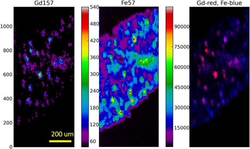
Gd and Fe (counts/s) in human liver imaged by LA-ICPMS with lateral distribution of 10 µm. Gd is accumulated in discrete areas, in some cases together with Fe (right image, colocalization areas are denoted with pink spots). Image credits: L. Pascolo, K. Vogel-Mikuš, J.T. van Elteren, N. Maximova. |
“We have found significant gadolinium retention in our child patients, even several months after MRI exams. We hope that the high chemical sensitivity and nanometric resolution of the ID16B beamline will enable us to unravel the mechanisms of hepatic Gd accumulation and the potential long term toxicity”, says Lorella.
“We are particularly interested in the relation between gadolinium and iron at intracellular structures. We are using the ESRF to understand where the gadolinium is lodged in the cells and to clarify any relation with the iron build-up”, adds Katarina Vogel-Mikuš from the University of Ljubljana Department of Biology in Slovenia.
The researchers hope to benefit from the unique opportunity offered by the advanced techniques available on ID16B synchrotron spectroscopy, nano-resolution imaging and high chemical sensitivity - to unravel the potential toxicity of gadolinium-containing MRI-CAs. The results could help them to understand the link between Gd accumulation and iron overload, opening a path to therapeutic options like chelating agents.
 |
|
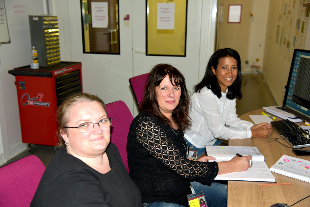
The team start to decipher the first results of scans on ID16-B. From left to right: Katarina Vogel-Mikuš, Lorella Pascolo and Vanessa Isabel Tardillo Suarez (ESRF). ©ESRF/K. Colvin |
Text by Kirstin Colvin
Top image: Inside the ID16-B experimental hutch, the team watch as the ESRF local contact, Vanessa Isabel Tardillo Suarez, explains parts of the set-up. Left to right: Lorella Pascolo, Anna Camillo, Vanessa, Alice Cernogoraz, Katarina Vogel-Mikuš. ©ESRF/K. Colvin
partners


European Synchrotron Radiation Facility - 71, avenue des Martyrs, CS 40220, 38043 Grenoble Cedex 9, France.