- Home
- News
- General News
- #weekendusers Getting...
#weekendusers Getting to the uncharted core of bones
26-06-2017
The skeleton is an essential part of our body and yet there is still a lot unknown about it. Bones can seem straight-forward to study, but their complex composition at the nanometre scale makes them a challenge. This weekend, scientists from Aarhus University in Denmark are delving into the core of bones on ID13.
When doctors diagnose diseases like osteoporosis, they generally base themselves on X-rays showing a smaller density of bones than what is normal. However, there can be individuals with dense bones that are still prone to unprovoked fractures and other with low density bones and with a low risk of fractures. Scientists are trying to get to the heart of bones, in order to understand, in the long term, how bone diseases appear.
Bone is formed primarily from collagen fibrils, calcium phosphate nanoparticles, water and non-collagenous macromolecules. It is traversed by a network of cells, called osteocytes, which are located in lacunae interconnected by canaliculi that are only a few hundred nanometre in diameter, the LCN. Osteocytes are constantly formed by the bone as it repairs itself.
The bone matrix around the LCN differs in structure and composition from the surrounding normal bone matrix and is enriched in non-collagenous proteins. “Today we have cartoon-like images on how bones look but there are disagreements in the community about the details”, explains Henrik Birkedal, the leader of the team. And he adds “The LCN makes up a sizeable portion of bone, yet almost nothing is known about the biomineral organisation or the distribution of oligo-elements in the bone matrix linked to the LCN”. This information is important to ultimately understand bone properties and the role of osteocytes, which are thought to play a key role in maintaining bone and controlling calcium levels in the body.
 |
|
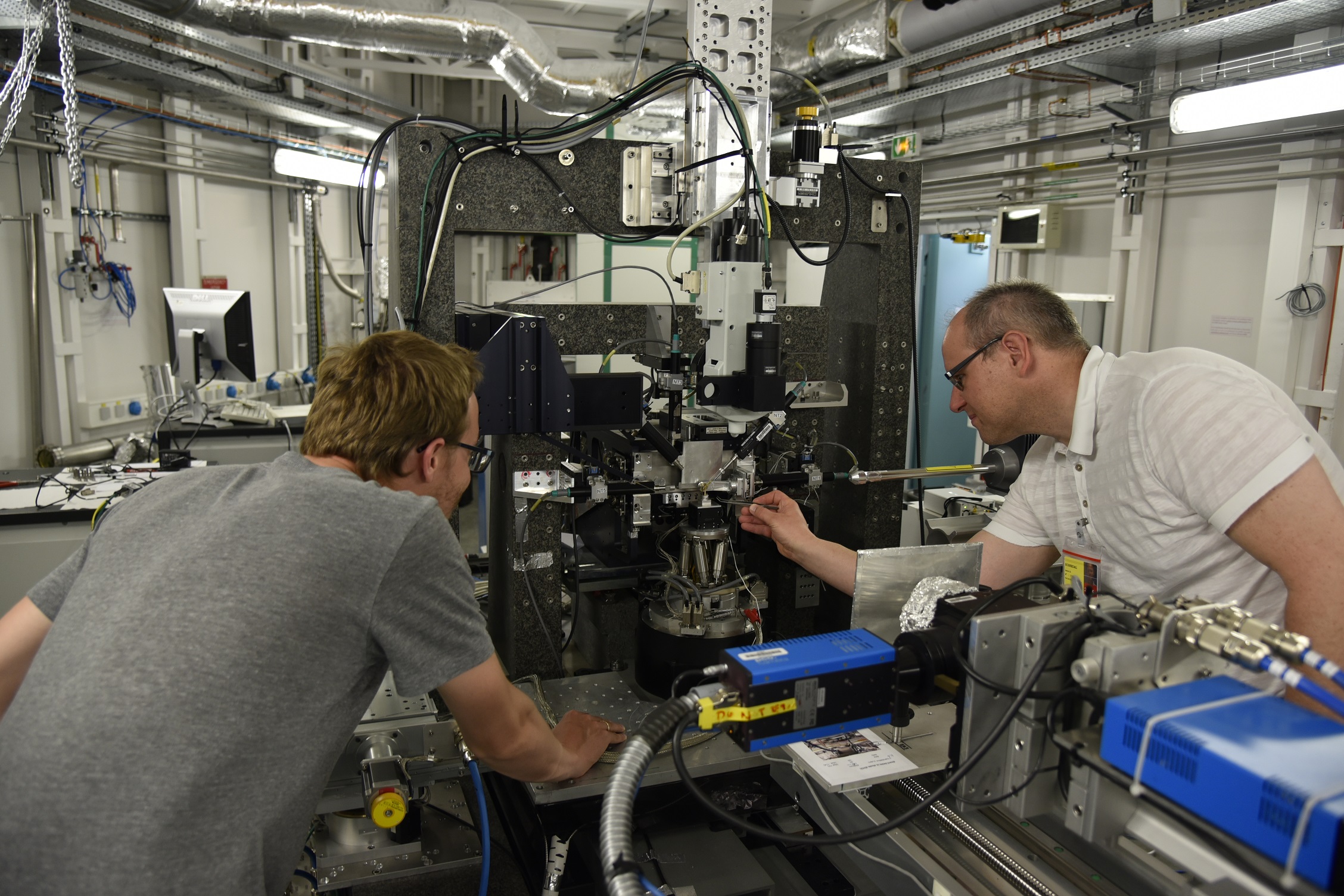
Placing the sample in the experimental hutch. |
Birkedal and his team will try to map the nanocrystals and oligo-elements by combining diffraction and fluorescence computed tomography on ID13. “We need beams that can go under 100 nanometres, as well as outstanding detectors such as what we have on ID13”, explains Birkedal. “We are actually attempting to break a resolution record, aiming for a 50-nanometre beam, so it is a challenging experiment”.
Getting down to this small beam sizes is not easy and it has been the subject of a long-term project collaboration between ID13, the Fraunhofer Institute of Materials and Beam Technology (Dresden, Germany) (Adam Kubec, Sven Niese) and the Erich Schmidt Institute of Materials Science (Leoben, Austria) (Jozef Keckes). The team very recently achieved a 35 nm beam by Multilayer Laue lenses, which will help researchers like Birkendal and his group to answer their scientific questions.
This is not the first time the Birkedal group is at the ESRF. Previous experiments on ID16 already concluded that there are additional void spaces in bone that were not previously accounted for.
 |
 |
|
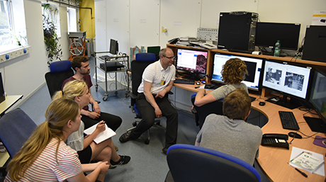
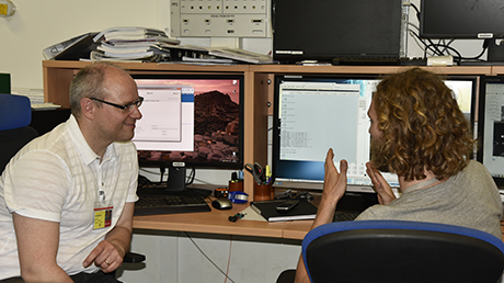
On the beamline's control hutch. |
|
The team is hoping to get a picture of the distribution of oligo-elements and calcium around the LCN (through fluorescence), as well as its mineral properties (through diffraction). The ultimate goal is to come up with improved models of bone’s hierarchical structure and osteocyte formation, which will help to understand bone mechanics.
 |
|
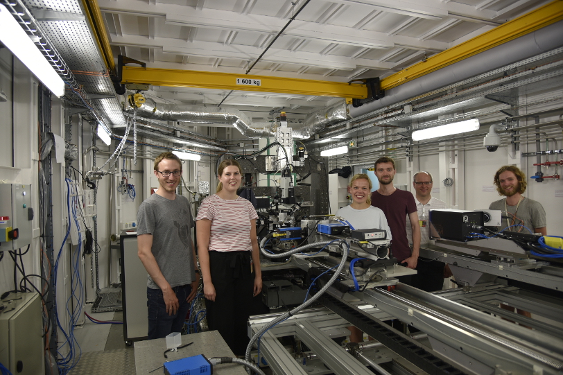
The team, from left to right: Morten Bormann Nielsen, Maja Østergaard, Nina Kølln Wittig, Jonas Palle, Henrik Birkedal and ESRF local contact Tilman Grünewald. |
Text by Montserrat Capellas Espuny
Top image: Placing the sample in ID13 experimental hutch
partners


European Synchrotron Radiation Facility - 71, avenue des Martyrs, CS 40220, 38043 Grenoble Cedex 9, France.