- Home
- Users & Science
- Scientific Documentation
- ESRF Highlights
- ESRF Highlights 2017
- Complex systems and biomedical sciences
- 3D quantification of vascular and neuronal alterations in a multiple sclerosis model
3D quantification of vascular and neuronal alterations in a multiple sclerosis model
X-ray phase-contrast tomography has been applied for the first time at micro- to nanoscales, with simultaneous imaging of vascular and neuronal networks, to obtain unprecedented direct 3D characterisation of the neuropathology in experimental autoimmune encephalomyelitis, the animal model for multiple sclerosis, and of the effect of mesenchymal stem cell treatment on tissue damage.
Multiple sclerosis is an autoimmune disease of the central nervous system associated with demyelination, axonal damage and neuronal loss resulting in neurological deficits. The use of appropriate animal models such as experimental autoimmune encephalomyelitis (EAE) facilitates the study of disease mechanisms and the development of new therapeutic approaches for multiple sclerosis.
Techniques previously used to investigate damage to vascular and neuronal networks in EAE suffer from several limitations. In particular, 2D imaging restricts spatial coverage, entails destructive sample preparation, and may lead to data misinterpretation due to lack of information in the third dimension. In contrast, recent work [1] on naïve mouse central nervous systems demonstrated that imaging by X-ray phase-contrast tomography (XPCT) enables the study of the 3D distribution of both the vasculature and single elements of the neuronal network, without sample sectioning and specific sample preparation.
Here, multiscale XPCT images have been generated to evaluate morphological alterations in vascular and neuronal networks at relevant disease phases of EAE in affected mice and to understand how treatment with mesenchymal stem cells (MSC) modifies them. A direct 3D morphological description of EAE lesions is provided at both vascular and neuronal levels at two different length scales. Micro-XPCT was performed at beamline ID17, giving a 3D quantitative analysis of the vasculature and of the neurons of the entire lumbar part of the spinal cord with spatial resolution of about 7 microns (voxel size of 3.5 micron), one day and five days post disease onset, without any staining procedure. Nano-XPCT was then performed at ID16A to investigate the 3D capillary network and nerve fibres in a region of about 300 x 300 x 70 mm3 with voxel size of 130 nm.
Such a multi-scale direct analysis has never been performed to understand EAE pathology and address the effect of an innovative therapeutic strategy.
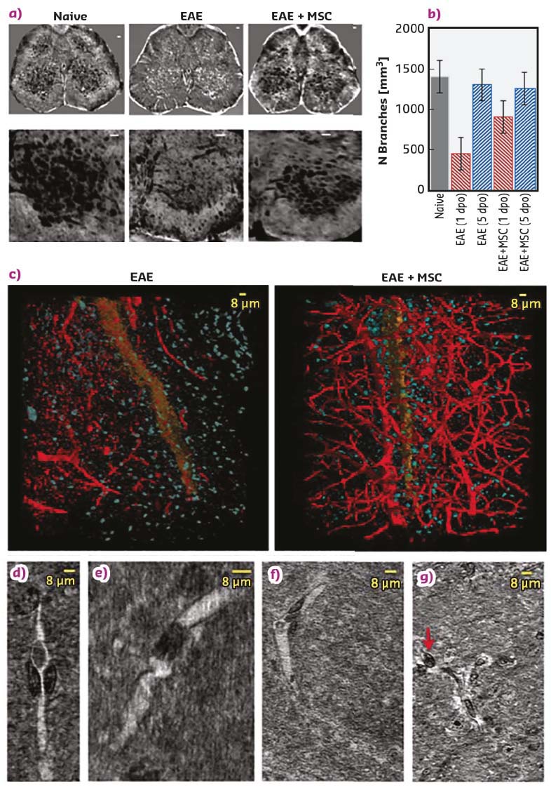
Vascular alterations have been described in EAE and multiple sclerosis, and analysis indicates that vasculature density is clearly decreased at disease onset in EAE-affected mice, a decrease not observed upon treatment with MSC (Figure 68), suggesting a protective effect of MSC on spinal cord vasculature. Most importantly, the XPCT study revealed an alteration of the capillary network in EAE (Figure 68) that had not been previously described. Thus, the results strongly indicate a trend in alteration of the 10-24 micron vessels and possible occlusions in the capillaries, an observation never obtained in tissue without the use of a contrast agent. Importantly, 3D analysis demonstrated that MSC administration reduces vascular alteration of vessels.
 |
|
Fig. 68: Vascular alterations in EAE redeemed through MSC treatment. a) Axial projections of XPCT images of spinal cord in naive, EAE-affected, and EAE-affected MSC-treated mice. Lower panels show segmentation of the vasculature (black) of the respective upper-panels. b) Quantification of the number of vessel-branches in lumbar spinal cord at one and five days post disease onset. c) Nano-XPCT volume renderings of the capillary network (red) and cell population (blue) in spinal cord of untreated and MSC-treated EAE-affected mice at five days post disease onset. The spinal canal is rendered in orange. Nano-tomography slices of capillary alterations in EAE (d-g). |
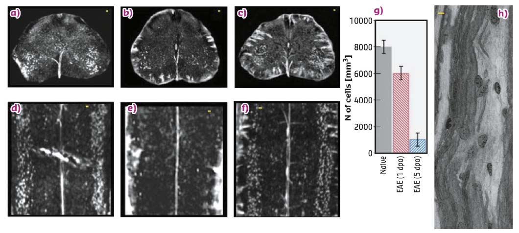
Simultaneous 3D XPCT imaging of neuronal alterations during EAE (Figure 69) supports the findings [2] of a massive loss of lower motor neurons in multiple sclerosis patients and in mouse EAE from early disease stages, with evidence of apoptotic neuronal death. The data suggest that the angiogenesis detected at the acute phase of EAE, by 2D immunohistochemical analysis of tissue slices, is in fact not efficient, and that the massive loss of neurons observed reflects the demonstration of pyknotic neurons in EAE observed by 2D techniques.
 |
|
Fig. 69: Effect of MSC treatment on the altered neuronal network in EAE (a-g), scale-bar 20 µm. Nano-XPCT (scale-bar 8 µm) of myelin damage in EAE (h). |
Through XPCT, never applied before to investigate multiple sclerosis models, crucial information not obtainable with other techniques has been derived, which can be used to further study disease evolution, as well as to optimise advanced new therapies and monitor their efficacy.
Principal publication and authors
X-ray phase contrast tomography reveals vascular alterations and neuronal loss in a multiple sclerosis model, A. Cedola (a), A. Bravin (b), I. Bukreeva (a), M. Fratini (a, c), A. Pacureanu (b), A. Mittone (b), L. Massimi (a), P. Cloetens (b), P. Coan (b, d), G. Campi (e), R. Spanò (f), F. Brun (a), V. Grigoryev (g), V. Petrosino (h), C. Venturi (h),
M. Mastrogiacomo (f), N. Kerlero de Rosbo (f) and A. Uccelli (f), Sci. Rep. 7, 5890 (2017);
doi: 10.1038/s41598-017-06251-7.
(a) Nanotec-CNR, Rome (Italy)
(b) ESRF
(c) Santa Lucia Foundation, Rome (Italy)
(d) Ludwig-Maximilians-Universität, München (Germany)
(e) IC-CNR, Rome (Italy)
(f) Department of Experimental Medicine, University of Genoa, Genoa (Italy)
(g) MEPhI, Moscow (Russia)
(h) DINOGMI, University of Genoa, Genoa (Italy)
References
[1] M. Fratini et al. Sci Rep. 5, 8514 (2015).
[2] J. Vogt et al. Ann Neurol. 66, 310-322 (2009).
partners


European Synchrotron Radiation Facility - 71, avenue des Martyrs, CS 40220, 38043 Grenoble Cedex 9, France.