- Home
- Users & Science
- Scientific Documentation
- ESRF Highlights
- ESRF Highlights 2016
- Structure of materials
- Getting to the heart of fossils
Getting to the heart of fossils
Most of the current knowledge on the evolution of vertebrates comes from fossilised bio-mineralised tissues such as bones and teeth. Very rarely, a fossil will present soft tissue preservation, allowing a leap in our understanding of evolution. This was the case for ca. 115 million year-old fish from Brazil, which were fossilised with their hearts.
The evolution of the chambered heart is a key point in the history of vertebrates. However, there have never been enough clues to elucidate this million-year-old process, since no proper vertebrate cardiac structures were ever described in the fossil record. This is partly because the heart is made from soft muscle tissue, which normally decays rapidly after death and is not preserved, unlike hard tissues such as bones.
The Santana Formation from the Brazilian Northeast is a geologic Konservat Lagerstätte (undisturbed sedimentary deposit), well known for its extraordinary fossils preserved in pristine conditions. From these ca. 115 million year-old sediments, the ray-finned fish Rhacolepis buccalis is known from hundreds of specimens, many of which are preserved in 3D in nodular concretions. Over the course of a wider study on potential heart fossilisation, a peculiar concretion was observed in one of these partially damaged fish: the location and shape of the concretion suggested it could have resulted from fossilisation of a heart.
Given the uniqueness that could represent the discovery of a fossilised heart, physical and chemical preparation techniques were excluded, favouring the non-invasive approach of X-ray tomography. To overcome the lack of contrast between this concretion and the surrounding sediment, the fossil was characterised using propagation phase contrast synchrotron X-ray microtomography (PPC-SRµCT) at beamlines ID17 and ID19, along with 60 other fossils of Rhacolepis from the same locality, holding hopes for other similar discoveries in unbroken specimens. On both beamlines, the specimens were characterised using high energetic beam (monochromatic on ID17, filtered white beam from a wiggler on ID19), using long propagation distances (ca. 11 m) and producing data with a resolution of 45 µm. Two of these additional fish showed structures that, when observed in reconstructed data, were undoubtedly identified as fossilised hearts. For these two, a second tomography was performed at 6 µm resolution.
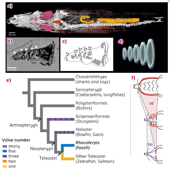
The heart remains were located posterior to gills and between the bones of the pectoral girdle, as in modern ray-finned fishes (Figure 62). Their configuration consisting of four distinct chambers (conus arteriosus, ventricle, atrium and sinus venosus), typical ventricular and atrial muscular trabeculae and paired Cuvier duct meeting the posterior-most chamber (sinus venosus). Within this architecture, special interest was given to the conus arteriosus, a conical extension of the ventricle that helps regulate blood outflow via valves.
 |
|
Fig. 62: Fossilised heart of Rhacolepis. a) 3D rendering of a complete Rhacolepis with various soft tissues preserved, including the heart (in red). b) tomographic slice showing the conus arteriosus in dark, partitioned by five valves (marked by white arrows). c) interpretative drawing of the tomogram. d) idealised, didactic model of the five valve system. e) simplified phylogeny of fishes showing the known number of valves observed in various lineages. f) Interpretative drawing illustrating the simplification of the heart architecture, from an outflow tract dominated by a multi-valved conus arteriosus (ca) at the top, through the only known fossil example with five valves, to the dominant, elastic, non-chambered, condition represented in the bulbus arteriosus in most modern fish (ba). |
Modern ray-finned fishes (actinopterygians) present different types of structure responsible for the outflow tract: in basal actinopterygians, such as the freshwater bichir Polypterus, the outflow tract is controlled by a dozen valves contained in the conus arteriosus. These valves prevent backflow and protect the delicate gill vessels from the elevated pulsation generated by the ventricle; in derived actinopterygians (e.g. the teleost zebrafish Danio rerio), this task is rather carried out by an additional cardiac segment, the bulbus arteriosus. This valveless structure protects the gills through its prominent elastic properties. The timing and details of this evolutionary transition in the heart architecture of fishes were almost completely unknown.
The fossilised heart of Rhacolepis displays a large conus arteriosus enclosing five conal valves. It contrasts with Polypterus in having a significantly reduced number of valves, the latter exhibiting nine valvar rows, each containing three to six individual valves. Rhacolepis also notably contrasts with the very limited number of valves observed in modern derived actinopterygians, which vary from one to two. Rhacolepis clearly shows an intermediary state between the ancestral multi-valvular condition toward a significant simplification, as known in many modern ray-finned fishes and suggests an overall gradual evolution, rather than an abrupt simplification.
Principal publication and authors
Heart fossilization is possible and informs the evolution of cardiac outflow tract in vertebrates, L. Maldanis (a,b), M. Carvalho (b,c), M.R. Almeida (d), F.I. Freitas (e), J.A.F.G. de Andrade (f), R.S. Nunes (g), C.E. Rochitte (h), R.J. Poppi (d), R.O. Freitas (g), F. Rodrigues (i), S. Siljeström (j), F. Alves Lima (g), D. Galante (g), I.S. Carvalho (k), C. Alberto Perez (g), M. Rodrigues de Carvalho (c), J. Bettini (l), V. Fernandez (m) and J. Xavier-Neto (b), eLife 5, e14698 (2016); doi: 10.7554/eLife.14698.
(a) Department of Pharmacology, University of Campinas (Brazil)
(b) Brazilian Biosciences National Laboratory, Campinas (Brazil)
(c) Department of Zoology, University of São Paulo (Brazil)
(d) Institute of Chemistry, University of Campinas (Brazil)
(e) Geopark Araripe, Crato (Brazil)
(f) National Department of Mineral Production, Crato (Brazil)
(g) Brazilian Synchrotron Light Laboratory, Campinas (Brazil)
(h) Heart Institute, InCor, University of São Paulo (Brazil)
(i) Institute of Chemistry, University of São Paulo (Brazil)
(j) Department of Chemistry, Materials and Surfaces, SP Technical Research Institute of Sweden (Sweden)
(k) Departamento de Geologia, Universidade Federal do Rio de Janeiro (Brazil)
(l) Brazilian Nanotechnology National Laboratory, Campinas (Brazil)
(m) ESRF
partners


European Synchrotron Radiation Facility - 71, avenue des Martyrs, CS 40220, 38043 Grenoble Cedex 9, France.