- Home
- Users & Science
- Scientific Documentation
- ESRF Highlights
- ESRF Highlights 2013
- X-ray imaging
- The interaction of asbestos and iron in lung tissue revealed by synchrotron-based scanning X-ray microscopy
The interaction of asbestos and iron in lung tissue revealed by synchrotron-based scanning X-ray microscopy
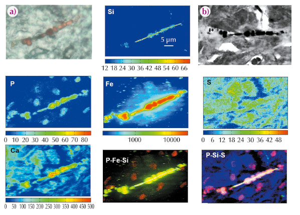
Asbestos is a potent carcinogen associated with malignant mesothelioma and lung cancer but its carcinogenic mechanisms are still poorly understood. Asbestos toxicity is ascribed to its particular physico-chemical characteristics including the presence of the fibres and their ability to adsorb iron, which may cause an alteration of the homeostasis of this element in the tissue. When asbestos fibres are inhaled, they may get trapped within the lung and remain indefinitely in the body locked inside biostructures called “asbestos bodies” which are the result of a complex biomineralisation process. Using a combination of advanced synchrotron-based X-ray imaging and microspectroscopic methods, we studied representative lung tissue samples from ten patients exposed to asbestos (from shipyard workers in Monfalcone, Italy). We obtained important correlative morphological and chemical information on the chemistry of asbestos body formation and other changes in the surrounding lung tissue. The iron concentration, distribution and speciation in diseased human lungs were analysed for the first time thanks to the high elemental and chemical sensitivity of synchrotron XRF spectro-imaging and micro-XANES available at beamline ID21. Our results contribute to an explanation of iron mobilisation while asbestos is present in lung tissue. Fe XRF maps (Figure 19, Fe panel) clearly demonstrate how the asbestos fibres and bodies can cause high mobilisation of iron into the surrounding cells (mainly alveolar macrophages) and tissue. The results suggest both continuous deposition of Fe-containing species (ferritin) around the asbestos fibres and metal release due to asbestos body degradation. Confirming and expanding on previous studies [1], the results showed that, along with iron, other chemical elements, including phosphorus, calcium and magnesium, participate in the formation of asbestos bodies and are also indicative of calcification processes. In Figure 19 the asbestos body is identified by light microscopy (panel a) as an orange-brown formation, while the X-ray absorption image in panel (b) (obtained at TwinMic beamline, Elettra Synchrotron) allows better definition of the features of the body. The XRF elemental maps allow identification of the original silicate fibre (Si map), from which the asbestos body originated. The coating is best represented by P and Fe maps. The Fe map clearly shows accumulation of Fe in the vicinity of the asbestos bodies, both as a diffuse signal and as bright spots resembling intracellular vesicles or siderosomes. Interestingly, P and S maps reveal cell structures: S seems to be a good marker of cell substance and useful to delineate cytoplasmic borders, while P seems to pinpoint the cell nuclei where the content of this element is high. When merging the P, Si and Fe maps (P-Fe-Si) or P, Si and S maps (P-Si-S), it appears that both the majority of iron diffuse signal and the vesicular spots, are mainly related to the presence of macrophage-like cells. The calcium distribution (Ca) appears to correlate with cell and tissue architecture but it is much more abundant in the coating of the asbestos body.
 |
|
Fig. 19: Micro-XRF and X-ray microscopy of lung tissue containing asbestos. (a) Visible light image; (b) X-ray microscopy absorption image (TwinMic beamline, 0.9 keV) and the corresponding Si, Ca, P, S and Fe XRF maps (ID21, 7.3 keV). |
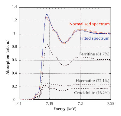
Since the oxidation state of Fe could provide important information on the processes involved in the asbestos body formation, Fe K-edge XANES spectra were collected in selected ~1 μm2 spots from the Fe maps at beamline ID21 (Figure 20). The measures confirm that most of the iron detected around asbestos fibres (coating and ferruginous bodies) is compatible with the presence of ferritin and the Fe3+ oxidation state of iron. In addition, micro-XANES analyses demonstrate their potential to discriminate the fate of iron-containing fibres from iron-free asbestos in lung tissues. However, the most novel and intriguing result of micro-XANES analyses was the detection of significant percentages of haematite in the asbestos bodies. We suppose that this is the result of ferritin transformation occurring during the prolonged residence time in the asbestos bodies in the lung tissues.
 |
|
Fig. 20: Micro-XANES analyses of asbestos bodies. An example of a deconvoluted micro-XANES spectrum of an asbestos body measured with a microprobe of 1 μm2 at ID21. |
Principal publication and authors
L. Pascolo (a), A. Gianoncelli (b), G. Schneider (c), M. Salomé (d), M. Schneider (e), C. Calligaro (f), M. Kiskinova (b), M. Melato (a) and C. Rizzardi (c), Scientific Reports 3,1123 (2013).
(a) Institute for Maternal and Child Health, IRCCS Burlo Garofolo, Trieste (Italy)
(b) Elettra - Sincrotrone Trieste S.C.p.a., Trieste (Italy)
(c) Department of Anatomical Pathology, University of Trieste (Italy)
(d) ESRF
(e) Hospital of Monfalcone, Monfalcone (Italy)
(f) Servizio Diagnostica Veterinaria, University of Udine (Italy)
References
[1] L. Pascolo, A. Gianoncelli, B. Kaulich, C. Rizzardi, M. Schneider, C. Bottin, M. Polentarutti, M. Kiskinova, A. Longoni and M. Melato, Part Fibre Toxicol. 8, 7 (2011).
partners


European Synchrotron Radiation Facility - 71, avenue des Martyrs, CS 40220, 38043 Grenoble Cedex 9, France.