Hardware
Hardware developements and sample preparation
Instruments are continuously developed at the beamline. For the study of artistic materials, a specific effort, made during the PhD of Emeline Pouyet was the assessment of micro-analysis of thin sections (instead of cross-sections) and the development of suitable protocols for sample preparation.
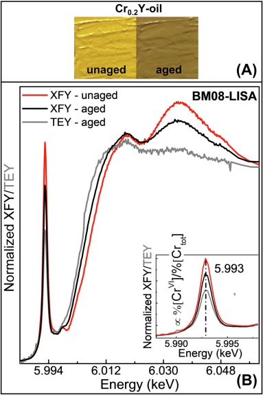 |
L. Monico, F. d'Acapito, M. Cotte, K. Janssens, A. Romani, G. Ricci, C. Miliani and L. Cartechini, “Total electron yield (TEY) detection mode Cr K-edge XANES spectroscopy as a direct method to probe the composition of the surface of darkened chrome yellow (PbCr1-xSxO4) and potassium chromate paints", Nuclear Instruments and Methods in Physics Research Section B: Beam Interactions with Materials and Atoms, (2023) |
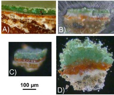
The darkening of chromate-pigments, including chrome yellows (PbCr1-xSxO4), is a surface phenomenon affecting late 19th-early 20th c. paintings, such as those by Van Gogh. Exploring analytical strategies that contribute to a deep understanding of darkening is therefore significant for the long-term conservation of unique masterpieces.
Here, we examined the capabilities of Cr K-edge XANES spectroscopy collected at the same time in X-ray fluorescence yield (XFY) and total electron yield (TEY) detection modes to selectively study the surface composition of darkened oil paint mock-ups composed of chrome yellow (PbCr0.2S0.8O4) or potassium chromate. By discussing advantages and drawbacks in using XFY/TEY modes in relation to XFY µ-XANES analysis from sectioned samples, we aim at assessing if TEY-XANES spectroscopy: (i) is a selective surface method to determine the abundance of different Cr-species from paint fragments; (ii) can contribute to optimize the analytical strategy by limiting time consuming sample preparation procedures; (iii) can decrease the probability of radiation damage.
Applications of FTIR to the study of ancient paintings have encountered two major steps: the development of FTIR microscopes, which enable to watch the sample and to select the regions of interest; and the migration of those FTIR microscopes towards synchrotron facilities, that offers a significant improvement in terms of spatial resolution and spectral quality. Even if access to synchrotrons is by nature limited, it is worth applying for such installations as they usually provide a set of micro-imaging techniques. FTIR end-stations should then be considered as part of a wider analytical platform. Combining micro FTIR with micro X-ray fluorescence, micro X-ray diffraction or micro X-ray absorption spectroscopy for the study of paintings enables a deeper insight into paintings at the micrometer scale. Several examples of X-ray/infrared combinations are given. The focus is given to practical aspects, in particular to the critical issue of sample preparation. Alternatives to the classical polished cross-sections are proposed and evaluated. Data treatment is also discussed.
|
E. Pouyet, B. Fayard, M. Salome, Y. Taniguchi, F. Sette and M. Cotte, "Thin-sections of painting fragments: opportunities for combined synchrotron-based micro-spectroscopic techniques", Heritage Science, 3, 1-16 (2015). |
|
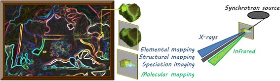
Synchrotron Radiation (SR) - based techniques such as SR-μ Fourier Transform InfraRed (FTIR) spectroscopy, SR-μ X-Ray Fluorescence (XRF), SR-μ X-Ray Absorption Near Edge Spectroscopy (XANES) and SR-μ X-Ray Diffraction (XRD) are increasingly used for the study of cultural heritage materials, as they offer enhanced spatial resolution and chemical sensitivity. For such analyses, painting fragments are usually prepared as thick (typically several hundreds of micrometers) polished cross-sections. The capabilities of these SR techniques can be significantly improved (enhanced data quality, reduced acquisition time, new imaging capabilities, improved lateral and in-depth resolution, reduced dose, etc.) if carried out on thin-sections, i.e. less than ~30 μm in thickness. This paper discusses and illustrates the different motivations in terms of related increased analytical capabilities for SR-μFTIR, SR-μXRF, SR-μXANES and SR-μXRD. Corollary to the optimization of the procedures for single SR micro-analytical technique, a specific discussion is focused on the challenges of their combination.
 |
E. Pouyet, A. Lluveras-Tenorio, A. Nevin, D. Saviello, F. Sette and M. Cotte, "Preparation of thin-sections of painting fragments: Classical and innovative strategies", Analytica Chimica Acta, 822, 51-59 (2014). |
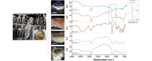
For more than a century, the analyses of painting fragments have been carried out mainly through the preparation of thick resin-embedded cross-sections. Taking into account the development of innovative micro-analytical imaging techniques, alternatives to this standard preparation method are considered. Consequently, dedicated efforts are required to develop preparation protocols limiting the risks of chemical interferences (solubilisation, reduction/oxidation or other reactions) which modify the sample during its preparation, as well as the risks of analytical interferences (overlap of detected signals coming from the sample and from materials used in the preparation). This study focuses particularly on the preparation of thin-sections (1–20 μm) for single or combined fourier transform infrared (FTIR) spectroscopy and X-ray 2D micro-analysis. A few strategies specially developed for the μFTIR analysis of painting cross-sections have already been reported and their potential extrapolation to the preparation of thin-sections is discussed. In addition, we propose two new specific methods: (i) the first is based on a free-embedding approach, ensuring a complete chemical and analytical neutrality. It is illustrated through application on polymeric design objects corpus; (ii) the second is based on a barrier coating approach which strengthens the sample and avoids the penetration of the resin into the sample. The barrier coating investigated is a silver chloride salt, an infrared transparent material, which remains malleable and soft after pellet compression, enabling microtoming. This last method was successfully applied to the preparation of a fragment from a gilded Chinese sculpture (15th C.) and was used to unravel a unique complex stratigraphy when combining μFTIR and μXRF.
 |
M. Cotte, J. Szlachetko, S. Lahlil, M. Salomé, V. A. Solé, I. Biron and J. Susini, "Coupling a Wavelength Dispersive Spectrometer with a synchrotron-based X-ray microscope: a winning combination for micro-X-ray fluorescence and micro-XANES analyses of complex artistic materials", Journal of Analytical Atomic Spectrometry, 26, 1051-1059 (2011). |
X-Ray fluorescence (XRF) is one of the most established techniques for the analysis of works of art. It offers several features—such as non-invasiveness, richness of information, in situ analysis with portable instruments and 2D and 3D micro-imaging when performed at synchrotron sources—which are increasingly used in the field of Cultural Heritage science. However, the complexity of artistic materials may lead into complex XRFspectra. Overlapping of fluorescence emission lines can make XRF analyses prone to error. In this article, we discuss the attributes of the polycapillary-based Wavelength Dispersive X-ray Spectrometer (WDS) developed at the X-ray micro-spectroscopy ESRF beamline, ID21. With spectral resolution of a few tens of eV, this system allows better separation of neighbouring emission lines. Its pros and cons for micro-XRF point analysis, micro-XRF mapping and micro-XANES are addressed, with a particular focus on Cultural Heritage applications. Possible extensions towards micro-RIXS are also discussed.
 |
M. Cotte, E. Checroun, V. Mazel, V. A. Solé, P. Richardin, Y. Taniguchi, P. Walter and J. Susini, "Combination of FTIR and X-rays synchrotron-based micro-imaging techniques for the study of ancient paintings. A practical point of view", e-PRESERVATIONScience, 6, 1-9 (2009). |
partners


European Synchrotron Radiation Facility - 71, avenue des Martyrs, CS 40220, 38043 Grenoble Cedex 9, France.