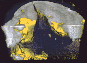
Figure 8

Fig. 8: X-ray phase-contrast tomogram using synchrotron radiation of a rat cerebellum. Grey and white matter and various substructures may be distinguished. Yellow: Sample surface structure. Violet: Embedding material. (Specimen provided by M.F. Rajewski, Institute of Cell Biology, University of Essen). The high sensitivity provided by phase contrast for imaging structural details in organic matter is evident. With absorption contrast practically no structures can be seen in this sample. Also of great importance is the much lower radiation dose deposited in the specimen when phase contrast is used.


