- Home
- News
- General News
- Size did matter
Size did matter
12-06-2009
Oldest evidence for reproduction with giant sperm uncovered at the European Synchrotron Radiation Facility
The mystery of giant sperm present in some living animal groups today has taken on a new dimension. In one group of micro-crustaceans new evidence shows the feature is at least 100 million years old.
Renate Matzke-Karasz, from Ludwig-Maximilians-Universität Munich (Germany), has led an international team of scientists, studying specimens from the London Natural History Museum’s collections. Their research has revealed fossilised evidence for reproduction using giant sperm in a group of small aquatic crustaceans, called ostracods, dating back to 100 million years ago.
Matzke-Karasz said, ‘In these microfossils, we detected organs that are required for transferring giant spermatozoa. Since modern ostracods still produce giant sperm and manoeuvre them with the same organs as 100 million years ago, it’s safe to say this distinctive feature evolved only once in this group. It seems to be an evolutionarily successful reproduction strategy, even though it comes at an exceedingly high price for both genders, as a lot of energy is invested in producing and carrying such enormous sperm.’
 |
|
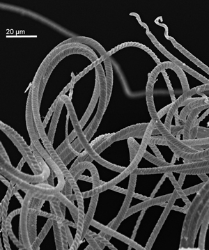
The length of the filamentous, helical sperm-cells of freshwater ostracods can reach up to ten times the body length of its producer. The longest known ostracod sperm cell is 1cm long. This scanning electron micrograph shows a bundle of such giant sperm extracted from Eucypris virens, which are approx. 1.8 mm long (Copyright: Dr. Renate Matzke-Karasz). |
The international team analysed Harbinia micropapillosa specimens from the Cretaceous Period that had remains of the soft body intact. These fossils had been collected, investigated and then donated to the Natural History Museum in 2000 by Robin Smith. Now at the Lake Biwa Museum, Japan, Smith is a member of the research team. Eight years later, the same specimens were analysed using synchrotron X-ray holotomography at the European Synchrotron Radiation Facility (ESRF) in Grenoble, through collaboration with Paul Tafforeau, a palaeontologist at the ESRF. This method is currently the most powerful and sensitive way to investigate in three dimensions and at a microscopic scale, the internal anatomy of exceptional fossils without damaging them. “Holotomography is a non-destructive imaging technique like computer tomography (CT), but we use powerful and coherent synchrotron X-rays leading to a sensitivity thousand times higher,” explains Paul Tafforeau of ESRF. “It is since very recently that palaeontologists use this technique to image fossils, but the results achieved so far show that this technique will surely lead to many important discoveries on fossils”, he adds.
The X-ray examination of the fossilised ostracods revealed direct parallels with the complex reproductive apparatus of modern relatives of these Cretaceous fossils. The team also came across something of a surprise: two of the female specimens had inflated cavities that only occur in modern ostracods that have recently mated, meaning fossil evidence for an insemination had been uncovered.
 |
|---|
|
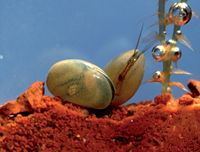
Mussel shrimps (Ostracoda) are small, bivalved crustaceans living in marine and freshwater environments. Their calcified carapace is readily fossilized, resulting in a fossil record stretching back almost 500 million years. This green freshwater species Eucypris virens prefers temporary pools (left in lateral view, right in ventral view, total body length approx. 1.8 mm) (Copyright: Dr. Renate Matzke-Karasz). |
The team was completed by Radka Symonová, scientist at the Charles University in Prague and Giles Miller, Micropalaeontology Curator at the Natural History Museum.
A human sperm would have to be over 17 meters long in order to measure up against one group of modern ostracods, whose sperm are up to ten times as big as the animals themselves. Roughly 34,000 of the 50 micron-long human sperm would have to line up to match the body length of a man (of 1,70m).
The next stage of the research from the international team is to understand why and how reproduction with giant sperm has persisted for so long.
Matzke-Karasz, R. et al, Science, 19 June 2009.
Notes for editors
• The research was led by Ludwig-Maximilians-Universität Munich and received funding from the ESRF (Grenoble), the European Union in the scope of the Marie Curie RT Network SEXASEX and the Lake Biwa Museum, Japan.
• Twenty-one specimens from the Natural History Museum’s micropalaeontology collection were used for the research and five of the specimens showed internal evidence of sexual organs
• The fossils were scanned using holotomography at the European Synchotron Radiation Facility in Grenoble, France, on beamline ID19
• All the holotomographic original and processed data presented in the publication will be made available to the public at the ESRF’s online palaeontology database, http://paleo.esrf.eu
• Modern animals that reproduce with giant sperm include featherwing beetles, several frogs, moths and backswimmer species, ostracods and fruit flies, such as Drosophila bifurca, whose sperm measures around six centimetres while the fly is only a few millimetres in length
Images:
EvirensLife.tif
Mussel shrimps (Ostracoda) are small, bivalved crustaceans living in marine and freshwater environments. Their calcified carapace is readily fossilized, resulting in a fossil record stretching back almost 500 million years. This green freshwater species Eucypris virens prefers temporary pools (left in lateral view, right in ventral view, total body length approx. 1.8 mm).
Copyright: Dr. Renate Matzke-Karasz
Sperm.tif
The length of the filamentous, helical sperm-cells of freshwater ostracods can reach up to ten times the body length of its producer. The longest known ostracod sperm cell is 1cm long. This scanning electron micrograph shows a bundle of such giant sperm extracted from Eucypris virens, which are approx. 1,8 mm long.
Copyright: Dr. Renate Matzke-Karasz
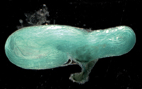
Receptacle.tif
Female freshwater ostracods store the giant sperm in two so-called seminal receptacles until they use the sperm cells to fertilize their eggs. When filled, these receptacles make up more than a third of the female’s body length. This picture shows a light microscopical view of an artificially stained receptacle of a Eucypris virens, filled to about half of its maximum capacity with sperm cells (receptacle length approx. 0.6 mm, sperm length approx. 1.8 mm).
Copyright: Dr. Renate Matzke-Karasz
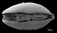
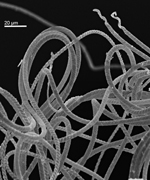
Harbinia.tif
Although the Cretaceous Santana Formation in Brazil is famous for its exceptionally preserved fish, it also yields ostracod fossils with preserved softparts – a very rare occurrence. The species Harbinia micropapillosa, here depicted as a scanning electron micrograph, reveals stunning details of the appendages. Now, a synchrotron holotomographic investigation showed that even internal organs can still be identified. The investigated fossils are housed by the Natural History Museum in London.
Copyright: The Natural History Museum, London
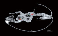
Tomography.tif
This synchrotron holotomography represents a virtual longitudinal section through the 100 million-year-old fossil ostracod Harbinia micropapillosa. The left arrow shows the preserved inner part of the oesophagus, while the right arrow points to the two seminal receptacles, where this female stored the giant sperm cells after mating.
Copyright: Dr. Renate Matzke-Karasz
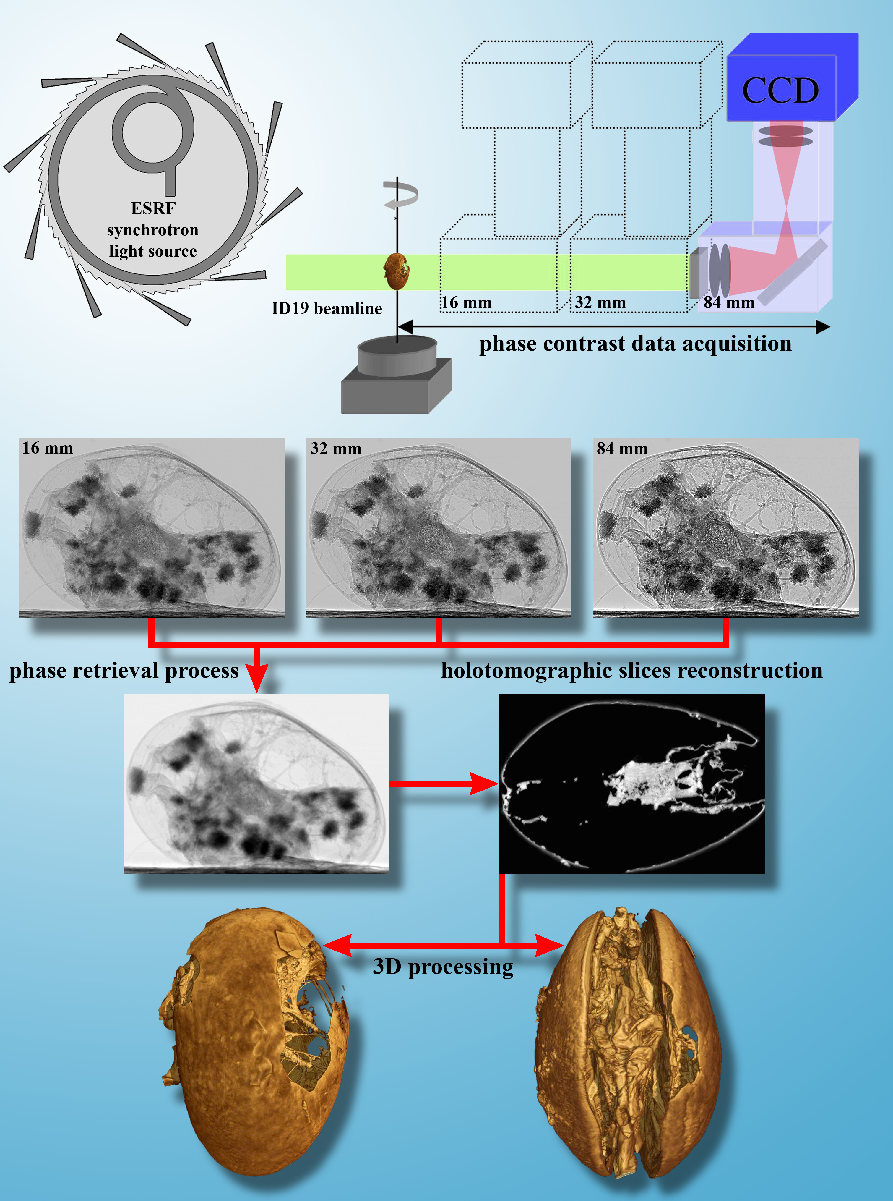
Holotomography.jpg
Principle of holotomography.
The study was mainly performed using holotomography. Holotomography (also known as inline quantitative phase contrast microtomography) is an X-ray imaging technique using the high coherence properties of X-ray beams produced by third generation synchrotron light sources such as the ESRF. It is based on several microtomographic scans (typically three or four) taken with increasing distances between the sample and the detector. During each scan, 1500 radiographs are taken during the rotation of the sample. When the sample detector distance is increased, it creates the “propagation phase contrast effect”, leading to an edge enhancement of all the structures of the samples which increases with distance. Combining these scans through a phase retrieval process allows the computing of a series of quantitative radiographic phase maps of the sample. These are directly linked to the densities of the sample, as in the case of classical absorption-based X-ray microtomography, but X-ray phase-contrast can be more than a thousand times more sensitive than X-ray absorption. The application of a tomographic reconstruction algorithm on the phase maps produces a holotomographic slice stack creating a 3D virtual volume of the fossil. The 3D processing and virtual dissections can then be made on these virtual holotomographic volumes.
Copyright: Paul Tafforeau (European Synchrotron Radiation Facility)
Videos:
15521_Xvid.avi
A 3D reconstruction of the ostracod derived from holotomographic measurements. To view these videos, you will need the open-source Xvid codec for Windows Media player, which can be downloaded here.
Copyright: Paul Tafforeau (European Synchrotron Radiation Facility)
Top image: A scanning electron micrograph of a Harbinia micropapillosa fossil. This millimetre-long creature is so well preserved that even the internal organs can still be identified.
partners


European Synchrotron Radiation Facility - 71, avenue des Martyrs, CS 40220, 38043 Grenoble Cedex 9, France.