- Home
- News
- General News
- New technique unveils...
New technique unveils the 3D composition of entire organic fossils
30-08-2019
Thanks to a technique developed at the ESRF, an international team of researchers has gained insight into the composition of a 53-million-year-old ant preserved in amber. The results are published in Science Advances.
An international team led by the IPANEMA laboratory (Paris-Saclay), and composed of 3 other CNRS labs (IMPMC, ISYEB, and LCPMR), the University of Lausanne and three synchrotron facilities (SOLEIL, the ESRF and SLAC) has established the chemical composition of a 53-million-year-old ant preserved in amber at a scale of one hundredth of a millimeter, using a new technique.
This technique, called 3D X-ray Raman imaging, has been developed at ESRF’s ID20 beamline. “We use hard X-rays but look at light elements, such as oxygen or carbon, and overcome the limitations of other techniques”, says Christoph Sahle, scientist in charge of ID20.
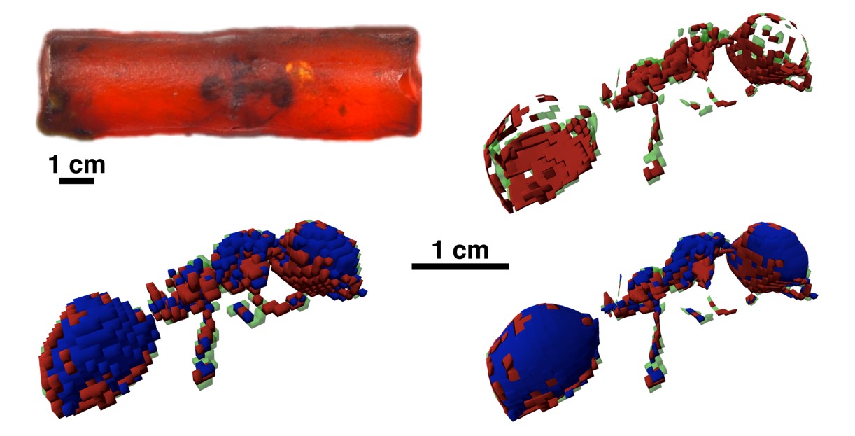
The results show the presence of molecular signatures of chitin, a complex sugar that constituted the insect's exoskeleton. They also reveal a difference in preservation between the part of the insect that was first in contact with the resin and the part that had been covered later, after the insect's death.
When using 3D X-ray Raman imaging, inelastic scattering transfers a small fraction of the X-ray energy with which a material is illuminated to its electrons, producing a detectable signal, used to determine its chemical composition. The new technique, in combination with the highly penetrating, focused, and monochromatic beam properties of synchrotron X-rays, allowed them to obtain the composition of the fossil in 3D. “This technique makes use of the innovative design of our new spectrometer, which allows us to explore spectroscopy in three dimensions”, explains Sahle.
 |
|
3D X-ray Raman imaging of an ant trapped in amber 53 million years ago in Oise (France). Carbon chemistry reveals the preservation of chitin molecular signatures, better preserved on the surface of the ant that was first in contact with the resin. |
For the last 15 years, the study of fossil materials has benefitted from 3D X-ray computed tomography, a method comparable to medical scanning. Tomography allows the study of microscopic patterns necessary to understand the evolution of species, their physiology and fossilization mechanisms. It leads to ‘black and white’ images indicative of the density of materials, in the same way that a medical X-ray displays the relative absorption of distinct tissues. Unfortunately, tomography does not provide the internal molecular composition of fossils, especially not of those composed of low Z-elements. Other methods, such as X-ray fluorescence imaging, can form a chemical image of high Z-elements, but only on flat fossils or thin sections.
“It is the first scientific case for this technique after five years of development, not only from the technical point of view but also from the data analysis side”, says Sahle. Loïc Bertrand, director of IPANEMA-CNRS and corresponding author, adds: "This collaboration, bringing together synchrotron facilities from around the world, illustrates the interest of seeking new and original methods for studying ancient and heritage objects, which have fundamental aspects still unknown to us”.
Reference:
Georgiou R. et al, Science Advances, 30 August 2019.
Text by Montserrat Capellas Espuny
Top image: Picture of one of the samples as it was placed in the experimental hutch during the experiment. Credits: A. Ciceron.


