- Home
- Users & Science
- Scientific Documentation
- ESRF Highlights
- ESRF Highlights 2006
- X-ray Imaging and Optics
- Non-destructive study of fossil inclusions in opaque amber using phase contrast X-ray synchrotron imaging
Non-destructive study of fossil inclusions in opaque amber using phase contrast X-ray synchrotron imaging
Amber is a plant fossil resin that dates from the Carboniferous period (~ 300/350 million years ago) to the present day. It sometimes contain fossil organisms that are generally in an excellent state of preservation. The study of these organisms permits a reconstitution of paleoenvironments and allows us to obtain pieces of information about the evolution of groups that are relatively rare in the fossil record, such as insects and spiders. Most well-known ambers originate from the Baltic Sea (Eocene, ~ 30-50 My) and from the Dominican Republic (Miocene, ~ 15-20 My).
During the last ten years, some sites with especially rich fossiliferous amber from lower Cretaceous (~ 100 My) have been discovered in Charente-Maritime (South-western France). These sites are studied intensively since lower Cretaceous sites containing amber are relatively rare. This period is also a key one in the Earth’s history with the explosion of phanerogams (plants with flowers), the maximum level of oceans and the maximum temperatures. Nevertheless, about 80% of the amber collected on these sites is opaque and cannot be investigated with classical optical methods, such as the optical microscope and binocular microscope, even after polishing.
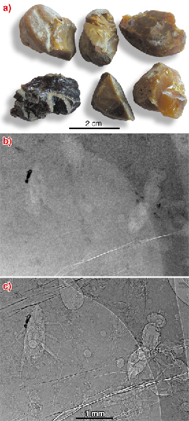
Finding the precise location of organisms embedded in the opaque amber was the first major problem encountered in this study. It was impossible to see anything using classical methods (Figure 141a), so synchrotron X-ray imaging was proposed as a solution for detecting the inclusions in amber [1]. Absorption radiography (Figure 141b) failed to reveal fossils in most cases due to their tiny volume, too small with respect to the volume of their containing amber blocs. Fortunately, propagation phase contrast was found to reveal the organisms very clearly (Figure 141c). This was initially demonstrated on isolated amber blocs [1], then the experimental protocol was optimised to permit a rapid survey of numerous amber blocs to find many fossil inclusions [2]. In only 48 hours, we succeeded in imaging about 2 kg of amber (corresponding roughly to a surface of 4000 cm2) with a pixel size of 5 µm, using propagation phase contrast radiography at beamline ID19. More than 350 well-preserved fossil organisms have been located and roughly identified (up to the family taxonomic level). This first survey showed a biological diversity as rich as the one observed in the small amount of transparent amber from the same sites.
 |
|
Fig. 141: a) Opaque amber blocs from Charente-Maritime. b) radiography of a bloc with inclusions in absorption mode. c) the same radiograph in propagation phase contrast mode with 990 mm of propagation distance (pixel size: 5 µm). |
 |
|
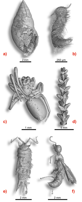
Fig. 142: Examples of virtual 3D extraction of organisms embedded in opaque amber. a) Gastropod Ellobiidae. b) Myriapod Polyxenidae. c) Arachnid. d) conifer branch (Glenrosa). e) Isopod crustacean Ligia. f) Insect hymenopteran Bethylidae. |
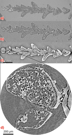
Some specimens were chosen for their preservation state and scientific interest, and scanned using phase contrast microtomography at beamlines ID19 and BM05 with various voxel sizes from 0.7 to 15 µm. Preliminary experiments demonstrated that that technique can reveal fossil insects in opaque amber in 3D with plenty of details [1]. 3D segmentation of the data allowed us to virtually extract the selected fossil organisms from resin in order to describe them in detail and to make a taxonomic identification of the species. More than 40 specimens have been scanned to date, and a diverse range of organisms has been discovered using this technique (Figure 142). The observation of microtomographic slices showed that most of the inclusions are empty. The original organism has completely disappeared, leaving an empty cast with all of the details, or, for some arthropods, an empty envelop corresponding to the original cuticle (exoskeleton). Sometimes, pyrite has crystallised in these cavities creating a replica of the organism. In a few rare cases, some internal organic structures are still preserved. For these particular specimens, we used holotomography (quantitative phase tomography) to enhance the visibility and contrast of preserved organic structures that are sometimes extremely tiny. For example, this technique permitted us to see cellular structures (cell walls and in some cases even the preservation of inner cell structures) on a small branch of a fossil conifer (Figure 143).
 |
|
Fig. 143: Comparison between scans in a) absorption mode, b) propagation phase contrast and c) holotomography, on the conifer branch (Glenrosa) with a voxel size of 15 µm. d) result of a high resolution holotomography (voxel size 1.4 µm) on the terminal burgeon showing clear cellular and tissue structures. |
This large survey of fossil organisms in opaque amber brings precious information about both the organisms and their ecosystem. The small gastropod (Figure 142a) indicates a close proximity of warm brackish water, the small isopod crustacean (Figure 142e) would have lived in a littoral area. These indications are in accordance with the hypothesis of a littoral environment like a lagoon or a mangrove swamp. Nevertheless, data are still insufficient from a statistical point of view to be able to obtain a reliable paleoenvironmental interpretation. This preliminary study was undertaken during a masters degree and concerned only 2 kg of opaque amber. There remains more than 80 kg to study, which should result in a very busy PhD thesis.
References
[1] P. Tafforeau et al., Applied Physics A, 195-202 (2006).
[2] M. Lak, Master2 dissertation (2006).
Authors
M. Lak (a,b), P. Tafforeau (b,c), D. Néraudeau (a), V. Perrichot (d) et A. Nél (e).
(a) Géosciences Rennes, UMR 6118, Rennes (France)
(b) ESRF
(c) LGBPH Poitiers, UMR 6046, Poitiers (France)
(d) Museum für Naturkunde, Berlin (Germany)
(e) MNHN Paris (France)
partners


European Synchrotron Radiation Facility - 71, avenue des Martyrs, CS 40220, 38043 Grenoble Cedex 9, France.