- Home
- Users & Science
- Scientific Documentation
- ESRF Highlights
- ESRF Highlights 2005
- X-ray Imaging and Optics
- X-ray Waveguides Deliver Ultra-small X-ray Beams
X-ray Waveguides Deliver Ultra-small X-ray Beams
The production of ultra-small and coherent hard X-ray nanobeams is currently an exciting field of research. Such beams would allow promising new applications such as holographic imaging and scanning X-ray probe experiments with unprecedented resolution. Among the different optics used to prepare X-ray nanobeams are Fresnel zone plates, curved mirrors, compound refractive lenses and X-ray waveguides.
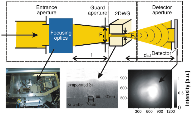
We have recently combined two independent optical elements for hard X-ray nanobeams (12.5 keV) to simultaneously achieve high gain and small cross section in two dimensions. An adaptive Kirkpatrick-Baez (KB) focusing optic [1] based on elliptically-curved mirrors was used to achieve a high flux density at the ID22 undulator beamline, with a focal spot of 2.5 x 3.8 µm2 (vert. x hor.) as measured by knife-edge fluorescence scans at the Au L-edge. This prefocused beam was coupled to the front side of a two-dimensionally confining X-ray waveguide (Figure 132). Depending on the lateral dimensions of the waveguide guiding core, the device allows the propagation of a defined number of modes. Owing to the filtering properties of the waveguide, the exiting beam exhibits a clean profile and it is not accompanied by any spurious reflected or transmitted beams which might present a source of complication in imaging or diffraction applications (Figure 133). Note that a mono-modal waveguide delivers a fully coherent beam (lateral coherence) [2].
 |
|
Fig. 132: Sketch of the optical system for nanobeam X-ray production. Left: The KB mirror optics is used to focus the beam. Middle: The waveguide filters the prefocused beam and further reduces its cross-section. Right: The exiting beam profile is characterised behind the waveguide. |
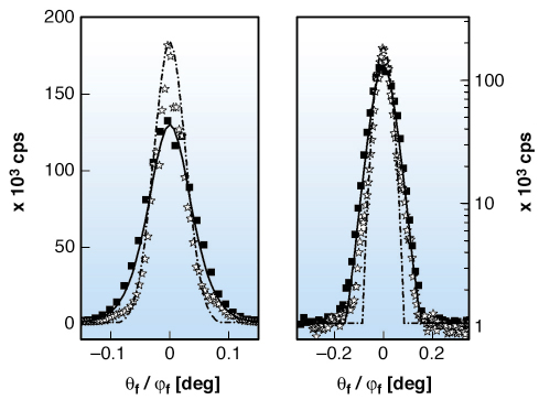
The waveguide was prepared by e-beam lithography. It consists of a polymer (PMMA) core (30 x 70 nm2 [vert. x hor.]) surrounded by silicon cladding on all four sides. The length of the device was 4.05 mm corresponding to an aspect ratio (length/width) of 60000. The high aspect ratio ensured negligible transmission of the primary beam through the silicon cladding (3.3 x 10⁻⁷ photons/s/mm2). The farfield pattern was measured by a CCD detector (Princeton Instruments) as well as by a scintillation detector for single photon counting (Cyber Star, Oxford Instruments). The vertical and horizontal line scans in the farfield (470 mm behind the guide) are in good agreement with the simulations of beam propagation (Figure 133). The total flux of the exiting beam was 3.5 x 10⁶ photons/s, corresponding to a waveguide efficiency of 4.7%, i.e. the number of photons exiting the device over number of photons impinging on the waveguide entrance. This corresponds to a overall system gain of g = 4000. Directly behind the guide, the beam has a lateral cross-section of 25 x 47 nm2 (Full Width at Half Maximum, FWHM), which is the smallest beam size for hard X-rays published so far.
 |
|
Fig. 133: The measured farfield beam profiles in the vertical ( |
Possible next steps include a better matching of the pre-focussing optics and the waveguides geometric acceptance. To this end, the KB mirrors could be placed at larger distances behind the source to further demagnify the beam. A reduction of the over-illumination at the waveguide entrance by two orders of magnitude thus seems to be feasible (thanks to the enhanced flux density). Finally, this non-dispersive optical system should allow the use of pink undulator beams, which could give an additional boost of another two orders of magnitude in coherent nanobeam flux.
References
[1] Y. Dabin, G. Rostaing, O. Hignette and A. Rommeveaux, Proc. SPIE, 4782, 235 (2002).
[2] C. Fuhse, A. Jarre, C. Ollinger, J. Seeger and T. Salditt., APL 85, 1907 (2004).
Principal Publication and Authors
A. Jarre (a), C. Fuhse (a), C. Ollinger (a), J. Seeger (a), R. Tucoulou (b) and T. Salditt (a), Phys. Rev. Lett. 94, 074801 (2005).
(a) Institut für Röntgenphysik, Göttingen (Germany)
(b) ESRF
partners


European Synchrotron Radiation Facility - 71, avenue des Martyrs, CS 40220, 38043 Grenoble Cedex 9, France.