Overview

From the very beginning the synchrotron radiation-based medical research programme has involved both imaging for diagnosis and irradiation for therapy, for pre-clinical and potentially clinical application.
A big effort was devoted to angiography, which aims at visualising coronary arteries. At beamline ID17 dual-energy images are acquired during the bolus passage of an iodine contrast agent after an intravenous infusion. At the moment studies have been performed on 51 volunteer patients (32 right coronary arteries, 19 left coronary arteries). This protocol should end up with 62 patients and might be continued by clinical trials like human kidney perfusion studies, follow up after arterial stent implantations or test of contrast agents.
Asthma is a disease from which more and more people suffer in our industrial societies. This motivated a programme to use the possibilities of synchrotron radiation to image the lung (Functional Lung Imaging Programme). Dynamic measurements of xenon concentrations in the airways are used to obtain maps of regional lung ventilation. We have studied local specific lung ventilation using tomographic and projection images. Recently, volume-rendering spiral computed tomography of rabbit lungs after an asthma crisis were performed. This tool is particularly valuable in the experimental study of small airway pathophysiology of the role of local mediators and that of pharmaceutical agents, and to develop experimental models of obstructive lung diseases such as asthma or emphysema.
Another disease which is very common and where early diagnosis is definitely very important is breast cancer. The Diffraction Enhanced Imaging (DEI) modality is especially interesting for radiation sensitive, soft-tissue imaging in mammography. The method is based on the use of highly-monochromatised radiation, passing through an analyser crystal in order to obtain an edge enhancement, at the interfaces of regions with different refractive indices. Excellent correspondence between tomographic slices and histological cuts of the same layer of cancerous tissue samples has been established. DEI offers a new kind of contrast for soft-tissue imaging based on the fact that eachè tissue species has its own characteristics with regards to small-angle scattering. Thanks to the low X-ray dose delivered in the experiments, a significant step towards clinical use of this method has been achieved.
Cerebral perfusion studies at the ESRF are made by temporal subtraction after the first passage of a contrast agent in the tomography mode. The accuracy and the precision of the synchrotron radiation computed tomography (SRCT) has been studied on the beamline. Direct measurement of the concentration of the contrast agent allows us to obtain functional parameters such as Cerebral Blood Volume or Flow and Permeability Coefficient (Figure 96). Such parameters are essential for the analysis of the physiopathology of brain diseases (mainly tumour processes), for therapy efficiency and for studying novel contrast agents issued from pharmaceutical companies.
The other important activity performed at the beamline is radiotherapy. Three radiation therapy techniques are currently under development on rats bearing cerebral tumours:
- Micro Beam Radiation Therapy is very promising for the treatment of cerebral tumours. It consists of a spatially fractionated beam, which allows the delivery of lethal doses to the tumour while sparing surrounding healthy brain tissues. Important steps from the pre-clinical phase towards the treatment of human tumours have been achieved.
- Another method is known as Tomotherapy. It consists of loading the tumour with a contrast agent in association with low energy X-ray irradiation (50-100 keV) during a rotation of the sample into the beam. The agent passes through the impaired blood brain barrier of the tumour vasculature. When using an iodinated contrast agent, the dose delivered to the tumour is enhanced thanks to the energy absorbed by the heavy element.
- Photon Activation Therapy is the irradiation of tumoural cells after introduction of cis-platinum in the cell culture medium. Cis-platinum is a widely used chemotherapy agent, it enters the nucleus and causes direct DNA damages when it produces photo-electrons under irradiation. Trials on rats bearing gliomas were carried out in various conditions of associated chemotherapy or contrast agent infusion and overall X-ray dose delivery. Outstanding increased survival rates were obtained with cis-platine infused directly in the tumour location before the irradiation in tomographic mode. Such results were the best ever obtained on pre-clinical trials aimed to cure the F98 model of brain glioma, and better than all others radiation therapy modalities. Moreover, basic studies dedicated to the analysis of the DNA damages and repair process on human cells incubated with cis-platinum showed the radiation damage depends on the wavelength when using a quasi-monochromatic beam.
The programmes developed on the ESRF medical beamline ID17 are the result of fruitful collaborations with different institutions located in different countries. The involvement of a team of the Grenoble university hospital allowed clinical protocols to became a reality.
Beamline characteristics
| wiggler on a low ß section | |
| critical energy | 33.5 keV |
| Kmax | 19.6 |
| field Bmax | 1.4 T |
| photon source divergence | 3.3 x 0.1 mrad2 (HxV) FWHM (at Bmax, 33 keV) |
| total power emitted | 14.3 kW (at Bmax) |
| Optics | ||
| optical elements | Si-bent Laue crystal | fixed-exit monochromator |
| distance from source | 144 m (approx.) | 140 m (approx.) |
| used for | angiography | computed tomography |
| spectral range | 17-80 keV | 15-80 keV |
| max beam size at patient | 150 x 10 mm2 (HxV) (aperture limited) | |
| expected flux | 2.1014 ph/s, 0.1% bw, 0.1 A, mradh, at Bmax, 33 keV,unfiltred | |
| Detectors | |
| angiography | Ge (2mm thick), 2 lines with 432 elements each (pitch 350 µm)-current integration mode |
| computed tomography | FRELON Camera with taper |
Preparation laboratory and Data Processing
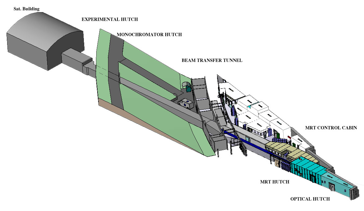
There are two medical research stations. An experimental station for radiotherapy is located in the experimental hall, 40 meters from the source point. Computed tomography and angiography take place in the satellite building, located 150 m from the source point.
Computing facilities are available to process images, CT reconstruction in 2D/3D. A DVD-R drive is available for data storage.
partners


European Synchrotron Radiation Facility - 71, avenue des Martyrs, CS 40220, 38043 Grenoble Cedex 9, France.