- Home
- Users & Science
- Scientific Documentation
- ESRF Highlights
- ESRF Highlights 2004
- Materials Science
- In situ X-ray Diffraction during Pulsed Laser Deposition
In situ X-ray Diffraction during Pulsed Laser Deposition
Pulsed Laser Deposition (PLD) has become a widespread technique for the fabrication of thin films [1]. A powerful pulsed laser is used to create a plasma off a target material, which is subsequently epitaxially deposited on a heated single crystal substrate. With each laser pulse, only a fraction of a monolayer is deposited, thereby making it possible to control the thickness very accurately. In between two individual laser pulses, the adatoms on the substrate are allowed to relax, which results in very smooth layers and interfaces. PLD is especially suited for the deposition of complex transition metal oxides and in particular for high-Tc superconductors owing to two different aspects. First of all, growth with PLD can take place at relatively high oxygen background pressures (up to 100 Pa), allowing for stoichiometric deposition of these materials. Second, the layered structure of high-Tc superconductors requires atomic control of deposition.
Although PLD is widely used due to the advantages over traditional molecular beam epitaxy (MBE) techniques, the processes governing the growth, i.e. nucleation and subsequent coalescence of islands are not yet understood. To accomplish a more detailed understanding of the PLD process, structural information during growth is needed. So far, mainly in situ Reflection High Energy Electron Diffraction (RHEED) measurements have been performed [2]. RHEED is a very useful technique to study the growth of the layer mechanism. However, a quantitative analysis of the structural data is hampered due to multiple scattering effects. In case of using X-rays, which interact much less than electrons in scattering events, kinematical theory applies. To study crystal growth using surface sensitive X-ray scattering techniques, high-brilliance X-ray beams, nowadays readily available at third generation synchrotron sources, are needed. In this way it is possible to investigate the growing interface/surface at an atomic scale.
To study thin film PLD growth in situ by X-ray diffraction, a special sample chamber was constructed [3]. A film of the high-Tc superconductor YBa2Cu3O7-x was grown on a substrate of SrTiO3. The experiments were carried out by mounting the chamber on a 2+3 type surface diffractometer with vertical scattering geometry at BM26 (Dubble) and focusing an excimer laser, having a wavelength of 248 nm, on the target. During deposition the substrate temperature was kept at 780C and the oxygen pressure in the chamber at 0.03 mbar.
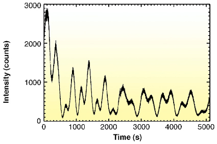 |
Fig. 28: Intensity oscillations of the (0,0,0.15) point as a function of time during growth of the YBa2Cu3O7-x thin film. |
At a grazing angle for the incoming beam of 1 degree, the intensity of the reciprocal point (0,0,0.15) in STO lattice units is monitored during deposition (Figure 28). An oscillatory pattern is seen where each maximum corresponds to the completion of another monolayer, and therefore indicating so-called layer-by-layer growth. This is explained by considering the following: on an atomically flat surface, a fraction of a monolayer is deposited. Due to the increase of the surface roughness, the intensity will drop. When however the monolayer is completed, i.e. the whole surface is covered by the monolayer, the surface is atomically flat again and the intensity reaches a maximum.
Growth was stopped several times, allowing further characterisation of the grown film. Figure 29 shows the 001 Bragg reflection of the film when it was 21.0(1) nm thick. The pronounced Kiessig fringes show that the surface is particularly smooth, which is to be expected in case of layer-by-layer growth.
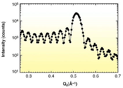 |
Fig. 29: The 001 Bragg reflection of the film at 21.0(1) nm thickness and 780 °C. Pronounced Kiessig fringes indicate a particularly smooth surface. |
In conclusion, we were able to follow the PLD growth of a thin YBa2Cu3O7-x film in situ by X-ray diffraction. This opens the possibilities of performing detailed studies of complex oxide thin film growth on the atomic scale.
References
[1] D. Chrisey and G. Hubler, Pulsed Laser Deposition of Thin Films (John Wiley and Sons, New York, 1994).
[2] G. Rijnders,G. Koster, D. Blank, and H. Rogalla, Appl. Phys. Lett. 70, 1888 (1997).
[3] V. Vonk, S. Konings, L. Barthe, B. Gorges and H. Graafsma, J. synchr. Rad. (in preparation).
Principal Publication and Authors
V. Vonk (a,b), K. Driessen (a), M. Huijben (b), S. Harkema (b), B. Gorges (a) and H. Graafsma (a).
(a) ESRF
(b) Low Temp. Division and MESA+ Res. Inst., Univ. of Twente (The Netherlands)
partners


European Synchrotron Radiation Facility - 71, avenue des Martyrs, CS 40220, 38043 Grenoble Cedex 9, France.