X-ray imaging
X-ray imaging techniques are used for innovative research in an ever broadening range of scientific areas. The variety is demonstrated by the articles in this chapter from the emerging field of palaeontology and of nano-imaging for biology and materials science. Also included are contributions on Biomedical applications, on Materials and Environmental science, and on Cultural Heritage.
Biomedical research is exemplified by three articles. The first is on the use of synchrotron radiation computed tomography for tumour functional mapping. This approach provides absolute values of different microcirculatory parameters for tumours and surrounding brain tissues. These measurements are of crucial importance for radiotherapy. The second article reports on functional imaging using the K-edge subtraction technique, here used to study the lung and bronchial response to methacholine treatment (a drug targeting lung function) in ovalbumin-sensitised rabbits. This work may have direct clinical implications and helps in furthering our understanding of asthma mechanisms and for tailoring drug-targeting strategies. The third article emphasises the importance of the nanoprobe beams produced at station ID22NI for biomedical purposes through a study of the metallobiology of the neuromelanin pigment in the human brain and its possible role in the pathogenesis of Parkinson’s disease.
X-ray imaging is widely used for Materials Science studies. The publications presented here take full advantage of the new capabilities of the imaging beamlines, i.e. fast 3D imaging and nanoprobe-related imaging. A full 3D microtomographic image can be recorded in only a few seconds on ID15 and ID19. This allowed in situ real-time studies of the dendritic solidification and coarsening in an Al–10 wt.% Cu alloy. The hard X-ray nanoprobe (ID22NI) was used to study the defects in GaN:Mg, and to produce sub-micrometre holotomography of multiphase metals. The following highlight describes the 3D investigation of cometary grains, collected during the NASA Stardust mission, by nanoscale X-ray fluorescence tomography.
Cultural Heritage studies and artefact preservation are increasingly important in Europe for both cultural and economic reasons. Synchrotron radiation has proven itself to be a valuable tool, allowing studies not otherwise possible. Over the past few years, Cultural Heritage related activities have undegone an extensive and successful development at the ESRF, as witnessed by the increase in the number of proposals to the Proposal Review Panel on Environment and Cultural Heritage (EC). X-ray imaging techniques play an important role in this area. This is exemplified by an article describing the full characterisation of a degraded cadmium yellow (CdS) pigment in an oil painting.
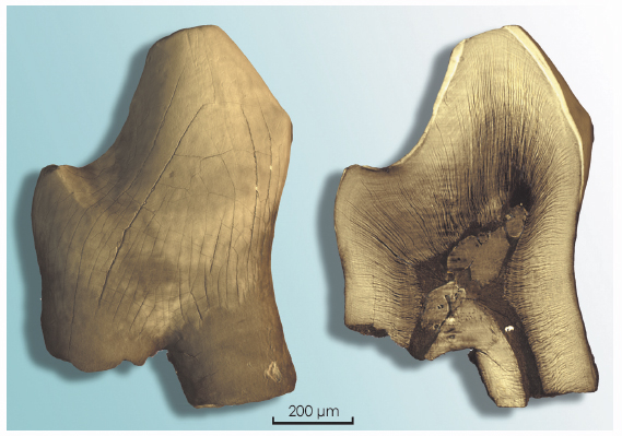
Within Cultural Heritage, we wish to emphasise the particular case of palaeontology. Since the first fossil was scanned at ESRF in 2000, palaeontology has become an important topic: the paleontological-related proposals have grown to about 20% of the proposals using microtomography. This research regularly leads to high impact publications (four in Nature/Science/PNAS in 2009) and to numerous PhD theses, and they have attracted substantial interest from the media and the public. In the present chapter, we present two publications performed using holotomography, the first on a very unusual (thought impossible) fossil fish brain, and the second to untangle the evolution of giant sperm in ostracod microcrustaceans. Another recent example is the sub-micrometre resolution holotomography of a mammalian tooth, presented in Figure 125, which reveals the internal histological information of dental microstructures, including the growth lines that constitute a record of an individual’s development.
 |
|
Fig. 125: 3D Rendering and virtual histological slice of a 145 million years old Cretaceous mammalian tooth from Cherves-de-Cognac (Charente, France). This minute fossil tooth was imaged using sub-micrometre resolution holotomography on ID19. The highly monochromatic source, in addition to short acquisition time, leads to unprecedented image quality for this kind of microfossil. The detailed 3D morphology gives access to the internal histological information of dental microstructures including its developmental record. Study of the growth lines in such fossils should bring new evidence of the controversial physiological thermal regulation in our mammalian ancestors as well as in dinosaurs (Courtesy: J. Pouech, J.-M. Mazin and P. Tafforeau). |
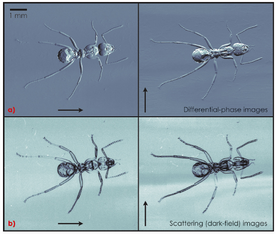
The scientific success of the X-ray imaging techniques would only be short-term without the constant effort of the ESRF staff and users to implement new imaging techniques that will lead to new scientific research. This is exemplified in the present chapter by a contribution on the extension of high-resolution laminography (which allows three-dimensional imaging of flat specimens) to weakly absorbing materials, through the use of propagation-based phase contrast. Another example is given in Figure 126, which shows a series of images providing complementary information recorded using a grating interferometric device. This phase-contrast technique can be applied to a wide variety of materials; it will be implemented for routine access for our users.
 |
|
Fig. 126: Radiographs of an ant taken with a two-dimensional X-ray grating interferometer (2D GIFM) with 23 keV X-rays. GIFM is a novel method that yields X-ray images with ultra-high sensitivity in several complementary contrast modes. With the standard interferometer, differential phase and dark-field images can be obtained along only one particular direction. An extended 2D version of the device has been implemented at ID19 permitting simultaneous access to the image signals along multiple directions. The two orthogonal sensitivity orientations of (a) differential phase contrast and (b) dark-field (scattering) contrast are indicated by arrows (Courtesy I. Zanette and T. Weitkamp). |
Several important publications have been produced by the two radiotherapy programmes under development at ID17: Stereotactic Synchrotron Radiation Therapy (SSRT) and Microbeam Radiation Therapy (MRT). These articles deal with the understanding of the response of healthy and tumoral tissues to X-ray treatment and with the refinement of clinical trials protocols for ID17.
X-ray imaging plays a substantial role in the ESRF Upgrade Programme, through which we intend to continue providing our users with the best instruments for their projects. A long (160 m) canted beamline at the ID16 location is planned for the ambitious Nano Imaging and Nano Analysis (UPBL4, NINA) project. A refurbishment of the parallel and coherent beam imaging beamline ID19 will also be implemented, with emphasis on palaeontological activities in close association with the Palaeontology Facility.
We are confident that these developments, coupled with the scientific know-how acquired using X-ray imaging at the ESRF, will allow the limits of the techniques to be extended, making possible scientific results which are not attainable today.
J. Baruchel
partners


European Synchrotron Radiation Facility - 71, avenue des Martyrs, CS 40220, 38043 Grenoble Cedex 9, France.