- Home
- Users & Science
- Scientific Documentation
- ESRF Highlights
- ESRF Highlights 2005
- X-ray Imaging and Optics
X-ray Imaging and Optics
Introduction
Synchrotron radiation based X-ray imaging and nanofocusing techniques are key elements in the preparatory document that lays out the long-term strategy of the ESRF. These techniques are promising for two reasons: firstly they are being applied to an increasingly large number of subjects including palaeontology (this Highlights cover) and environmental sciences (see Figure 131). Secondly, the evolution of these techniques towards quantitative measurements with high spatial and temporal resolution opens new prospects in research including the flourishing area of nano-technologies.
The development of SR-imaging and nanofocusing techniques depends on several technological improvements, particularly in the fields of mechanical and thermal stabilisation, and X-ray Optics, as shown in the first section of this chapter, X-ray Imaging Methods and Instrumentation. Two approaches are used to focus the beam to a nano-spot: the combination of an adaptive Kirkpatrick-Baez (KB) focusing optic based on elliptically-curved mirrors with X-ray waveguides, on ID22; and dynamically-bent graded multilayers, on ID19. The new sources will surely require optics elements able to withstand high heat-loads. Emphasis has therefore been put onto the collaboration with diamond producers, to obtain the highest quality diamond crystals. Some of the samples obtained to date already approach the required quality, as shown by the topographs. The two other contributions within this section describe ways to enhance the SR-based imaging techniques to offer improved opportunities for fundamental and applied science. The first involves free-space propagation and analyser-based imaging for phase contrast imaging. The second presents the results of a recently-designed laminograph, which permits three-dimensional images of flat samples that cannot be visualised using classical microtomography.
The two following sections are devoted to the application of X-ray imaging techniques to life science and medicine, on the one hand, and to the study of materials, on the other. The mechanical feasibility of a more advantageous interlaced cross-firing geometry for microbeam radiation therapy, and the validation of the use of monochromatic quantitative computed tomography for a direct measurement of the concentration of a contrast agent, are demonstrated. The application of X-ray imaging to materials is exemplified by two very different topics, the microtomographic study of a coronary stent, to reduce the risk of lesions, and results that shed light on an old but unresolved problem, the mechanisms of liquid metal embrittlement.
 |
|
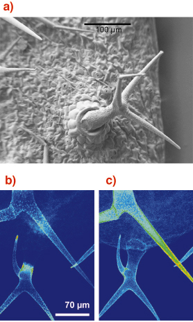
Fig. 131: (a) Scanning electron microscope image of "trichomes" from the plant Arabidopsis thaliana. (b) Fluorescence image showing the cadmium content. (c) Fluorescence image showing the calcium content. Images courtesy of M.-P. Isaure, B. Fayard, G. Sarret, S. Pairis, J. Bourguignon (Publication submitted to Environmental Science and Technology) |
The last section, X-ray fluorescence and diffraction microscopy, shows the same wide variety of topics, which range from the conservation of Henry VIII’s warship Mary Rose, to the critical fluctuations near a phase transition in Fe3Al, through the investigation of extraterrestrial grains for the NASA Stardust Mission, and finally to the evidence of diffusion at room temperature in a high Tc superconductor.
We wish to illustrate this dynamically-growing area of microspectroscopy with a recent environmental science application: the chemical images of small "trichomes" on plants leaves (see Figure 131a). Metal accumulation occurs in these epidermial hairs covering the leaves. It is well known that cadmium is highly toxic for plants: its distribution was studied by µXRF on ID21. The metal was found to be concentrated in localised areas of the trichomes (Figure 131b), which were also found to be enriched with calcium (Figure 131c).
J. Baruchel
partners


European Synchrotron Radiation Facility - 71, avenue des Martyrs, CS 40220, 38043 Grenoble Cedex 9, France.