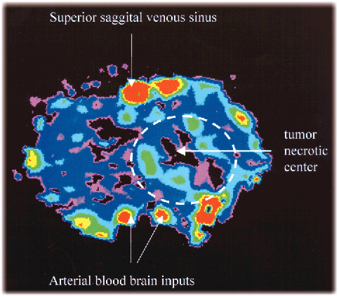
Figure 23
Fig. 23: Cerebral blood volume of a rat brain bearing a C6 glioma (axial orientation) obtained after a bolus infusion of 0.1 ml iodine based contrast agent (350 mg.ml). Slice thickness 0.35 mm, pixel size 0.35 x 0.35 mm2, slice level 9 mm above the line between the external auditive meatus. Colour table from red to black (100%, 0% respectively). High vascular density is visible surrounding the necrotic centre of the tumour.
| back to: Cerebral Blood Volume Measurements in Bolus Method with Synchrotron Radiation Computed Tomography |


