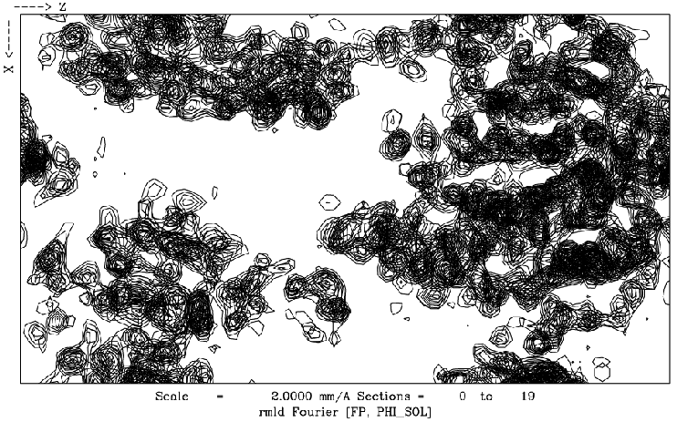
Figure 13
Fig. 13: Part of an electron density map of the protein L-rhamnose (J. Naismith and colleagues, St Andrews, Gordon Leonard, ESRF and Sean McSweeney, EMBL). This map was computed only some 50 minutes after the mounting of the sample on the beamline. It is clearly interpretable in terms of the molecular structure.
| back to: Multi-wavelength Anomalous Dispersion (MAD) Studies |
partners


European Synchrotron Radiation Facility - 71, avenue des Martyrs, CS 40220, 38043 Grenoble Cedex 9, France.