Figure 7
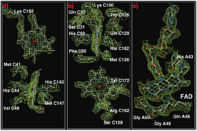
Fig. 7: Representative parts of the experimental electron density maps for crystal form A calculated with the MIRAS phases after density modification and phase extension to 2.2 Å resolution. Carbon, nitrogen, oxygen, phosphorous, and sulphur atoms are shown in grey, blue, red, light green and green, respectively, haem iron centres are shown in orange. Contour levels are 1.0 ![]() (green) and 9.0
(green) and 9.0 ![]() (red) above the mean density of the map. a, b) the two haem b molecules and the side chains of some neighbouring residues in the transmembrane region. c) the covalently bound FAD prosthetic group.
(red) above the mean density of the map. a, b) the two haem b molecules and the side chains of some neighbouring residues in the transmembrane region. c) the covalently bound FAD prosthetic group.
| back to: Fumarate Reductase from Wolinella Succinogenes |
partners


European Synchrotron Radiation Facility - 71, avenue des Martyrs, CS 40220, 38043 Grenoble Cedex 9, France.