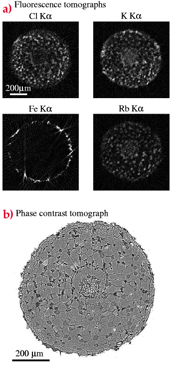
Figure 6
Fig. 6: (a) Fluorescence tomographs of the root of the mahogany plant (Swietenia macrophylla). Tomographic reconstructions (using filtered back projection) of the K![]() -lines for Cl, K, Fe, and Rb are shown. (b) Phase contrast tomograph of the mahagony root, showing the cellular structure.
-lines for Cl, K, Fe, and Rb are shown. (b) Phase contrast tomograph of the mahagony root, showing the cellular structure.
| back to: In vivo Fluorescence Microtomography of Plants |
partners


European Synchrotron Radiation Facility - 71, avenue des Martyrs, CS 40220, 38043 Grenoble Cedex 9, France.