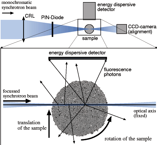
Figure 5
Fig. 5: Schematic setup for fluorescence microtomography. The sample is scanned through the microbeam for a large number of rotation angles between 0 and 360°. The fluorescence radiation is collected in the energy dispersive detector.
| back to: In vivo Fluorescence Microtomography of Plants |


