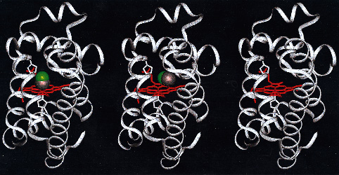
Figure 107
Fig. 107: Snapshots of the transition from MbCO to Mb obtained from 150 picosecond Laue data. The first picture shows the initial state before the optical pulse where the CO, shown in green and black, is bound to Fe in the center of the heme. The second picture is taken 4 nanoseconds after photon dissociation. It shows an intermediate state in which CO is rotated 90° and displaced 4 Å from Fe. It stays in this configuration ca 350 nanoseconds. The function of the docking site is to prevent the CO from recoiling back to the highly chemically attractive Fe. The last picture, taken 1 microsecond after the optical pulse, shows that the CO has left the pocket. It diffuses about in the outer protein for a fraction of a millisecond and comes then back to the Fe by random collisions with atoms fluctuating inside the heme pocket.
partners


European Synchrotron Radiation Facility - 71, avenue des Martyrs, CS 40220, 38043 Grenoble Cedex 9, France.