- Home
- Users & Science
- Find a beamline
- Structural biology
- How to use our beamlines
How to use our beamlines
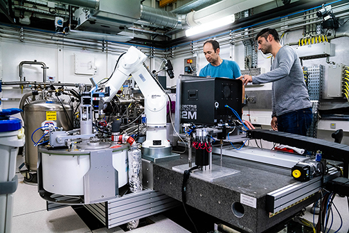
These page contain all the information required to complete a successful experiment at the Macromolecular Crystallography Group beamlines of the ESRF.
Before you come to the ESRF please read the "Prepare Your Experiment" pages. Once here the "Run Your Experiment" pages will help you along the way and if you get into any difficulties the "Trouble Shooting" pages should have the answers. All of our beamlines are equipped with a high precision minidiffractometer and a sample changer (for which you must use SPINE standard pins).
Please cite the paper describing MXCuBE for experiments conducted at the ESRF Structural Biology Beamlines, Gabadinho, J., et al. (2010).MxCuBE: a synchrotron beamline control environment customised for macromolecular crystallography experiments. J. Synch. Rad., 17, 700-707
partners


European Synchrotron Radiation Facility - 71, avenue des Martyrs, CS 40220, 38043 Grenoble Cedex 9, France.